Retinal detachment after surgery is a very common phenomenon that can manifest itself in absolutely no way, especially at the beginning. For this reason, patients require a visit to an appropriate specialist for diagnosis, as well as a thorough examination of the fundus of the patient. Detachment is dangerous, first of all, due to the fact that under high voltage this process can accelerate significantly and become a direct cause of a sharp deterioration in vision. Later stages are fraught with the appearance of myopia, in such situations, patients begin to "fly flies" in front of their eyes.
The operation, according to reviews, with detachment of the retina, is usually carried out using extrascleral filling and laser coagulation. It happens that it is necessary to completely or partially remove the vitreous body - vitrectomy.
Indications for surgery
Operational measures should be taken in case of retinal detachment. In a similar situation, two layers are separated - pigmented and neuroepithelial. Between these tissues, fluid usually accumulates. The filling is intended to restore the integrity of the membrane, and also helps to restore the lost functions to the eyes.
With minor damage, as well as with the preservation of vision and peripheral exfoliation, coagulation is done. Existing gaps on the background of this remain, but seem to be sealed around the edges. As a result, stratification does not spread, nor does a person's vision deteriorate. Laser surgery for retinal detachment is very popular.
The vitrectomy procedure is carried out when detecting changes in the vitreous, which is a gel-like substance that fills a significant part of the eyeball. Such an operation can also be prescribed for extensive damage to the retina, bleeding in the cavity of the vitreous body, as well as pathological vascular germination. According to reviews, surgery for retinal detachment is not suitable for everyone.
Contraindications for surgical measures for retinal detachment
Each of the above types of surgery has its own contraindications, so vitrectomy is not performed in a number of the following cases:
- With significant changes in the cornea and retina. In such a situation, surgery will not have the desired result.
- With significant opacification of the ocular cornea. They are usually visible with the naked eye in the form of a thorn.
Extrascleral filling is prohibited when:
- The extreme opacity of the vitreous.
- With ectasia or, in a more understandable language, protrusion of the sclera.
Laser coagulation should not be done with:
- Fundus hemorrhage.
- Opacity of the eye.
- High degree of retinal detachment.
- Pathology of the vessels of the iris.
How does the operation go when detached retina? About it further.
Among other things, contraindications remain when there are prohibitions on anesthesia and allergies to anesthetics. The operation cannot be performed in the presence of active inflammation. That is why, before going for surgery, it is necessary to pass all the necessary tests, fluorography, and in addition, you should get rid of tooth decay. The reasons for retinal detachment after surgery are of interest to many.
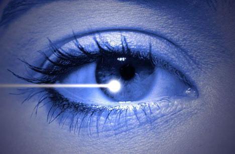
Laser coagulation as a method of surgical intervention
The operation cannot be performed without anesthesia. The whole process lasts about five to ten minutes. As a rule, in private clinics this procedure is not accompanied by hospitalization, and patients can leave the medical institution directly on the day of the correction itself. In public hospitals, the situation is somewhat different and patients are required to be monitored for three to seven days after surgery when the retina is detached. Reviews confirm this.
Since the procedure itself cannot be performed without anesthesia, only a small amount of anesthetic is used in the form of eye drops. In addition, doctors use drugs that are aimed at dilating the pupils. Immediately, from the moment their action begins, a special lens is put on the patient’s eye, which resembles the eyepiece of a microscope. This tool makes it possible to focus the laser beam and directs it to the desired location. During the operation, areas of protein destruction and the so-called gluing of the retina are created, which prevents its further separation.
How is the operation?
The procedure is carried out in a sitting position. The patient feels the laser in the form of bright light flashes. In rare cases, they can cause nausea and dizziness. For prevention, the patient is advised to concentrate all his attention on the second eye. During surgical operations, a slight tingling sensation is possible. Adhesions are finally formed after ten days or two weeks, and already after this period it will be possible to judge the success of the operation. How is recovery after surgery with detachment of the retina, consider below.
Extrascleral Filling Details
Before this operation, the patient should strictly observe bed rest. At rest, the liquid that is in the stratification zone is absorbed, and the emerging bubbles become more distinct. In the framework of extrascleral filling, such measures help to accurately determine all places of ruptures.
At the first stage of the surgical intervention, the surgeon cuts the conjunctiva, that is, the outer ocular membrane, and causes pressure on the sclera through a special device called a diathermocauter, which is an apparatus with various tips that allow you to create an electrical discharge of the desired intensity directly on the surface of the tissue. As a result of the above actions, a temporary shaft is created - the place where the sclera is pressed against the retina. This, as it were, marks all the places of delamination, after which individually a seal of the required size is made. Rehabilitation after surgery for detached retina is extremely important.
Then apply a soft and elastic material, usually silicone. A seal is placed on the sclera - the shell, which is located under the retina. As a result, all layers are pressed against each other, and the work and functioning of the visual apparatus are gradually restored. Sew the seal with threads that are not able to resolve. The fluid in the gap is gradually absorbed due to the pigmented epithelium. It happens that with its excessive accumulation, incisions are made in the sclera for complete elimination.
In some situations, the retina can additionally be pressed on the other hand, as if from the inside of the eye. For this, air is pumped into the vitreous, or another gas mixture. In this case, the patient is required to look in a certain direction and lower his eyes down. This allows the gas bubbles to fit exactly at the point of rupture. In order to replenish the volume, it is possible to introduce an isotonic solution into the vitreous body. The conjunctiva is sutured.
Operation success
Even despite the high degree of complexity of laser surgery for detachment of the retina, its success is quite high. In the textbook entitled "Eye Diseases" V.G. Kopaeva, which was released several years ago, said that when performing surgery using the modern technical level, it becomes possible to achieve retinal fit in ninety-five percent of patients. At the moment, surgical professionalism has noticeably increased, and the equipment used has become even more affordable and perfect. An important circumstance is the current diagnosis, which is carried out during periodic examinations by an ophthalmologist.
Vitrectomy with retinal detachment
This operation is carried out in stationary conditions. It is usually a complement to extrascleral filling, if there are appropriate indications. Vitrectomy is done both under general anesthesia and with local application.
Small holes are made in the sclera, into which tweezers and thin scissors are inserted. The vitreous is completely or partially removed, and the space that is freed is filled with silicone oil or a gas mixture.
Possible complications and consequences, based on medical data and patient reviews
In their reviews, the following processes are called the most common negative consequences after surgery:
- Some write that after surgery they develop strabismus. This complication is observed in half of those who have undergone extrascleral filling. Strabismus begins as a result of muscle damage during surgical intervention, as well as due to intergrowth of muscles with the sclera and so on.
- Many patients experience a change in vision. At first, the operated eye does not clearly perceive the contours of objects, so people most likely will need glasses with different diopters during the first few months. You have to visit an ophthalmologist from time to time to regularly check visual acuity. But, according to experts, after a few months, usually all indicators return to normal and stabilize.
- Inflammation, which manifests itself in itching, redness of the eyes, and tearing. As part of treatment and prevention, doctors advise the use of drops containing an antiseptic, which should be used within seven to ten days.
Recovery period
When performing laser coagulation, almost no restrictions are imposed on the patient. Patients can only be recommended exercises that should be aimed at strengthening the motor muscles of the eye. The doctor will probably advise you not to resort to strong and debilitating physical exertion during the first time after the procedure.
The speed of patient rehabilitation varies depending on the intensity of the regeneration of the body, the starting area of damage, as well as the degree of surgical intervention. As a rule, it lasts from ten days to several months.
We examined how the operation goes when retinal detachment.
Instead of a conclusion
Reviews of surgery for detachment of the retina in most cases are positive. The recovery period passes quickly, there are practically no complications.