It happens that the picture in front of my eyes suddenly blurred. Colors become less bright, objects lose their clarity, the world around us is plunged into "fog". Veil in the eyes - a fairly common phenomenon, but, alas, not harmless. Thus, the body signals serious ailments that could lead to loss of vision. Especially dangerous are the conditions when the veil does not look like a thin film, but like a cloudy, dark or red glass. Such a symptom warns of a violation of the transparency of the ocular media or problems of recognition of the received image by the occipital cortex.
Where to go
If you have a veil in your eyes, blurry vision, then first of all you need to contact an ophthalmologist. It is this specialist who must conduct the initial diagnosis and determine the cause of the disease. If the ophthalmologist does not find abnormalities, then a neurologist consultation will be needed. The main thing is not to delay the appeal, because you can lose time.
White shroud. Cataract
White veil in the eyes is most often a symptom of eye diseases. In many cases, this can be a one-way process involving only one eye. Often, patients with such complaints are diagnosed with cataracts, that is, a violation of the transparency of the lens.
The lens is a “biological lens” created by nature to refract light. It is located on the ligaments inside the eye and does not have its own blood supply. Nutrition of the lens is provided by intraocular fluid. At some point, as a result of natural aging or metabolic disorders, the transparency of the lens deteriorates. At this moment, a veil appears in the eyes, blurry vision, objects begin to double, obsessive flies appear in front of the eyes, the picture turns yellow, it becomes more difficult to read, write and work with small objects.
A person does not feel pain in cataracts, this creates a deceptive feeling that nothing supernatural is happening. However, the quality of life is gradually deteriorating, twilight vision is weakening, lacrimation begins in bright light, it is more difficult to read, more powerful lamps are needed, a halo is visible around the light sources, and patients with hyperopia gradually stop using glasses.
Glaucoma
Permanent veil in the eyes can be a symptom of glaucoma. This disease is associated with a steady increase in intraocular pressure, as a result of which intraocular hypertension begins, since the unhindered outflow of intraocular fluid is disrupted. The process is very dangerous, it can lead not only to impaired vision, but also to its complete irreversible loss. Suffice it to say that out of the total number of blind people, 15% lost their vision as a result of glaucoma.
Glaucoma is divided into two forms:
- Open angle. This means that the outflow of fluid in the anterior ophthalmic chamber in front of the lens has been impaired. Such a pathology is considered less dangerous, since it develops gradually, leaving time for action. With the open form of glaucoma, the angle of view gradually decreases (with a separate speed for each eye), a veil appears in the eyes and rainbow circles in front of them. Headaches become more frequent, twilight vision worsens.
- Angle angle. This means that outflow blocking occurred in the area where the iris and cornea join. At this point, the main fluid exchange of the anterior and posterior ophthalmic chambers occurs. At the first stage, the disease does not cause discomfort. The reasons for the deterioration of vision are not clear to the patient. Then an acute attack occurs during which the outflow of fluid is completely blocked. There is a sharp pain in the head and eye, which is often confused with migraine. Vision drops rapidly, a veil appears, dizziness and vomiting begin. The eye, in which an attack of angle-closure glaucoma occurred, turns red and becomes dense. Nature took very little time to eliminate outflow blocking. Sometimes it is only 3-4 hours. Then vision is lost forever.
Optic neuritis
As already mentioned, if there is a veil in the eyes, the reasons do not always lie in the field of ophthalmology. If, as a result of the inflammatory process, the optic nerve reduces sensitivity, the image from the retina does not reach the brain. This problem is called "optic neuritis" and is treated by a neuropathologist. In addition to inflammation, a cause of neuritis can be a demyelinating disease (destruction of the myelin sheath of neurons by the immune system).
The veil in the eyes, the causes of which are optic neuritis, may be accompanied by partial or complete blindness. The severity of the disease depends on the degree of damage to the diameter of the nerve.
A few more reasons for the white veil
In addition to the above diseases, the appearance of a white veil in front of the eyes can be caused by:
- occlusion of the central artery in the retina;
- corneal disease;
- presbyopia;
- brain tumor;
- uncontrolled intake of glucocorticoid drugs, antidepressants, contraceptives;
- dry eye syndrome.
Dark veil. Migraine
The veil before the eyes may not be whitish, but dark. This symptom is characteristic of several diseases, one of which is migraine. In this case, the causes of visual impairment are neurological in nature and are accompanied by a painful unilateral headache. Most often, patients have a genetic predisposition to migraine. Attacks of pain cause not only blurred vision, but also dizziness, nausea, speech impairment, and sometimes even hallucinations.
Retinal disinsertion
This is a problem associated with detachment of the inner lining of the eye containing photoreceptor cells. The retina at the place of detachment does not receive nutrition from the choroid, and dries out. The process is gradual, it begins with light flashes, zigzag lightning and black “flies”. Then a partial or complete dark veil appears in the eyes. What to do in this case? Urgent run to the doctor! Slight peeling can be “soldered” without serious consequences. But if the process is running, then the shrunken retina cannot be secured. Vision will be lost.
Red veil in the eyes
And another dangerous symptom is a red veil. This means that blood has poured into the vitreous body or the surrounding space, that is, hemophthalmus has occurred. In this case, the veil in the eyes may indicate a complication of diabetes mellitus, the development of atherosclerosis, hypertension or retinal degeneration. Hemophthalmus can also occur due to retinal detachment and eye injuries of varying degrees of complexity.
The shroud of red color is accompanied by blurred vision, the appearance of shadows, flies or stripes. If hemorrhage occurs as a result of glaucoma or trauma, then an additional symptom will be pain.
Why is it important to correctly describe the accompanying symptoms?
To make a correct diagnosis, it is not enough for the doctor to hear from the patient: "I have a veil in the eyes in the morning." A description of the accompanying symptoms will help you understand which direction to move. So, for example, if the appearance of the veil is accompanied by weakness, then it can be low blood pressure, anemia, VVD, hypertensive crisis. If random flies appear along with the veil, then cataracts, hemophthalmus, retinal detachment, a brain tumor (in the occipital part), migraine and others are added to the list of possible diseases. If dizziness is added to these symptoms, then it can be a stroke, internal bleeding, cerebral atherosclerosis, poisoning, and so on.
Since there are many variants of diseases, it is necessary to describe your condition to the doctor as accurately as possible.
Diagnostic examination
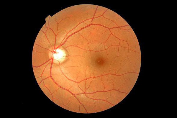
Since patients are primarily referred to an ophthalmologist, an examination will be carried out using a slit lamp, tonometry of the eye (measurement of intraocular pressure), hardware examination of the fundus, ultrasound. If the ophthalmologist does not detect pathology, the patient is redirected to a neurologist.
Neurologist determines reflexes and sensitivity, prescribes dopplerography of blood vessels (head, neck), MRI (head, neck).
Veil in the eyes: treatment
There are many factors that lead to vision problems. And each disease, a symptom of which could become a veil in the eyes, requires appropriate treatment. So, for example, with retinal detachment, drug therapy is prescribed that improves vascular patency and metabolism. Additionally, laser coagulation (soldering) of the retina is performed.
With cataracts, in its initial stage, vitamins and nutrients are prescribed for instillation into the eyes. At later stages, the lens is replaced.
With glaucoma, drugs are prescribed that reduce the pressure inside the eye. If necessary, the outflow is restored by surgical intervention.
The main thing that the patient should understand: the doctor needs time to take measures to preserve his vision. The shroud in the eyes cannot be ignored, especially if it is often repeated or held steady.