Visual impairment in hypertension is a secondary phenomenon. It is associated with changes in blood vessels. The degree of damage to the organs of vision can be different and manifests itself in the form of edema of the optic nerve, hemorrhages, detachment, retinal necrosis and other dystrophic processes. The eyes, along with the kidneys, brain, and blood vessels, are the target organs that are most affected by hypertension.
Eyes - a mirror of cardiovascular pathologies
According to various experts, a change in ocular day with hypertension is observed in 50-95% of patients. Periodic examinations by an ophthalmologist are one of the mandatory types of diagnostic tests for such patients. Monitoring the status of target organs is carried out for such purposes as:
- determination of the prognosis of arterial hypertension (AH);
- control over the course of the disease and visual impairment;
- assessment of the effectiveness and safety of therapeutic methods.
In modern international guidelines for the management of patients with arterial hypertension, a system of criteria characterizing the risk and degree of damage to various organs in hypertension is constantly updated and developed. Especially important are changes in the fundus with hypertension in the initial stages of this disease, since the deterioration is often asymptomatic.
The blood supply to the optic nerve inside the orbit is via the posterior ciliary arteries. The retinal central vein provides blood circulation in the retina. Disruption of blood flow under the influence of adverse factors leads to a decrease in metabolism in the retina and optic nerve.
Classification
Changes in the fundus with hypertension passes through several stages (Keith-Wagner classification):
- Broken or segmental, mild narrowing of small blood vessels and arteries. Lack of hypertension (high blood pressure).
- Stronger vasoconstriction, retinal vein displacement to its deeper layers, cross-linking with arteries due to arterial wall pressure.
- Damage to the retina due to severe vascular disorders (its edema, small and large hemorrhages, the appearance of bloodless foci such as "cotton spots"). The general condition of the patient is characterized by impaired cardiac and renal activity, high hypertension.
- Deterioration or complete loss of vision due to severe narrowing of the arteries and arterioles, edema of the retina and optic disc (optic nerve disc), the appearance of solid exudates around it. Severe condition of the patient.
This classification was first proposed in 1939 and is currently the most common in medical practice. At the same time, it was proved that the state of the fundus vessels in hypertension is a prognostic parameter of the lethal outcome in patients with hypertension. The disadvantages of this classification include difficulties in determining the initial stage of damage to the retina (retinopathy), the lack of a clear relationship between the stages and severity of hypertension. Some signs may develop inconsistently, which is associated with individual characteristics of the blood supply to the organs of vision.
The occurrence of retinopathy
Changes in the fundus with pressure are due to the following mechanisms:
- Short-term narrowing of small blood vessels at the initial stage as a result of the launch of the mechanism of blood flow autoregulation. Increased blood speed as a result of increased pressure. Changes in vascular resistance as a result of the body's adaptive ability to maintain stable blood flow.
- Thickening of the inner layer of arteries and veins due to chronic increase in vascular pressure, active neoplasm of smooth muscle fibers and destruction of fibrillar protein. Generalized narrowing of the small arteries.
- With the growth of destructive processes, large molecules penetrate from the blood vessels into the retina, the cells of the smooth muscle tissue and the layer lining the arteries die. The blood supply to the retina is significantly impaired.
Diagnostics
Fundus examination for hypertension is carried out by two main methods:
- Ophthalmoscopy - examination with an ophthalmoscope, which is included in the standard diagnosis by an ophthalmologist
- Fluorescence angiography. Before the procedure, a special substance is introduced intravenously - sodium fluorescein. Then a series of shots are taken when irradiated with a light source, as a result of which this compound begins to emit electromagnetic waves. Normally, the dye does not penetrate beyond the vascular wall. If there are defects, then they become visible in the picture. The duration of the procedure is about half an hour.
In older people over the age of 65, hypertensive syndrome can be mistakenly established, as retinal hemorrhages and fluid secretion through blood vessels are often caused by other causes. According to some reports, the diagnosis of hypertension, made according to the results of an ophthalmological examination, is true only for 70% of patients. In the late stage of the disease, the absence of specific changes in the retinal vessels is observed only in 5-10% of patients.
Differential diagnosis during the examination of the fundus for hypertension is carried out with such pathologies as:
- diabetes;
- consequences of radiation exposure;
- obstruction of the lumen of the veins and carotid artery (ocular ischemic syndrome);
- connective tissue diseases.
A key sign of hypertensive retinopathy is a change in blood pressure.
Description of the fundus in hypertension
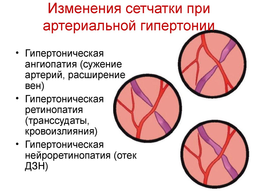
In ophthalmology, 2 types of changes in the fundus are distinguished - with and without retinopathy. In the first case, the initial transformations of the vascular network are observed, the arteries are still rectilinear, but their walls are already becoming dense and press on the veins, reducing their clearance. With a prolonged condition, retinopathy occurs, complicated by hemorrhages and exudate secretions from small arteries.
In the fundus with hypertension, the following pathological processes occur:
- angiopathy;
- arteriosclerosis;
- retino- and neuroretinopathy.
Patients with high blood pressure may develop retinal infarction, which leads to irreversible visual impairment. The inner surface of the eye normally looks as follows:
Photo fundus with hypertension, depending on the nature of the lesions, is presented below.
Changes in blood vessels
At the bottom of the eye, 2 vascular trees stand out: arterial and venous, which are characterized by several parameters:
- severity;
- branching and its features;
- the ratio of diameters (normal arteriovenous ratio is 2: 3; with hypertension, it decreases);
- sinuosity of branches;
- light reflex.
With hypertension, arteries often become less “bright”, the pattern of blood vessels becomes poorer (the same phenomenon is observed with myopia). This is due to a decrease in blood flow intensity. With increasing age, the arterial tree also appears less noticeable due to compaction of the vessel wall. Veins acquire a darker color and are better visualized. In some patients with good vascular elasticity, plethora is observed in both the arterial and venous trees.
Narrowing of the arteries during examination of the fundus with hypertension, is observed in only half of patients. It may have the following features:
- asymmetry of arteries in the right and left eye;
- uneven cross-section of one artery in the form of a chain of clamps and enlarged sections;
- Change only individual branches.
In the initial stages of hypertension, this is due to the uneven contraction of blood vessels in different areas, and during the period of sclerotic changes, when functional tissues are replaced by connective tissue, due to local thickening of the walls of the vessels. Prolonged hypertension leads to chronic retinal hypoxia, impaired function, protein dystrophy.
Mutual arrangement
One of the common symptoms of angiopathy is a violation of normal branching and the location of blood vessels in the fundus with hypertension. In a healthy person, the arteries are divided into two equal branches, which diverge at an acute angle. In patients with hypertension, this angle is increased (a sign of "bovine horns"). This is due to increased pulse blood strokes. An increase in the divergence angle helps to slow down blood flow in this area, which leads to the following negative consequences:
- sclerotic changes;
- blockage of blood vessels;
- destruction of the arterial wall due to lateral and longitudinal extension.
One of the most important and most common diagnostic signs of fundus disorders with hypertension is the intersection of arteries and veins, called the Hunn-Salus symptom. However, this phenomenon is also characteristic of arteriosclerosis without hypertension.
In this case, venous blood vessels are squeezed. This phenomenon develops in 3 stages:
- narrowing of the diameter of the vein under the artery;
- squeezing the vessel and its displacement deep into the retina;
- complete venous compression, lack of visualization of a blood vessel.
Retinal arteriosclerosis
Typical symptoms of retinal damage in hypertension associated with retinal arteriosclerosis are as follows:
- The appearance of light bands along the vessels (in ophthalmology they are called "cases"). This phenomenon is associated with a thickening of the vascular wall and a deterioration in its translucency.
- A wider and less bright reflex on arterial vessels.
- The syndrome of "copper wire" (a yellow tinge, detected mainly on large branches) and "silver wire" (a bright white reflection, which most often occurs on small arteries, the diameter of which does not exceed 50 microns).
The appearance of a light reflex along the vessels is explained by sclerotic changes in them, impregnation of their walls with exudate, as well as deposits of fat-like substances. At the same time, the vessels become pale and seem empty.
Hemorrhage
Hemorrhages in the fundus with hypertension appear due to the following reasons:
- leakage of blood cells through the violated vascular barrier;
- rupture of the aneurysm (the place where the artery wall is stretched and it protrudes) under the influence of high blood pressure;
- microthrombosis.
Most often, they appear near the optic disc in the form of radially directed strokes, “tongues of flame” and stripes. In the central region of the retina, hemorrhages are also located radially to the periphery. Less commonly, hemorrhages form in the layer of nerve fibers in the form of spots.
Exudates
Another sign of negative changes in the fundus with hypertension is exudates of gray-white color, soft, friable consistency, reminiscent of cotton wool. They develop rapidly over several days, but do not merge with each other. At their core, these formations represent a heart attack of a layer of nerve fibers that occurs due to the deterioration of blood flow in the blood vessels. There is a violation of the connection between the body of the neuron and its end. Nerve fibers swell and then break down. These necrotic processes are characteristic of other pathologies:
- diabetic retinopathy;
- blockage of the lumen of the central retinal vein;
- congestive optic disc disease, or swelling of the eye disc in the absence of inflammation, which occurs due to a slowdown in the flow of fluid from the eyeball to the brain (this condition can occur with changes in intracranial pressure).
The structure of solid exudates in the retina of the eye includes fats, high molecular weight proteins, the remains of cells and macrophages. These formations can be of various shapes and sizes. Their appearance is associated with the penetration of blood plasma through the walls of small blood vessels and the degeneration of surrounding tissues. Exudates can spontaneously resolve within a few months if there is a tendency to improve the condition.
Edema formation
The occurrence of retinal edema and optic nerves in the fundus with hypertension indicates a malignant course of hypertension. Accumulation of edematous fluid due to circulatory disorders leads to an increase in protein content. As a result, the retina becomes opaque.
Edema of the optic nerve can be in various forms - from mild to the development of congestive optic disc syndrome with hemorrhages, exudates in the central zone of the retina and foci of local heart attack.
The combination of signs of angiopathy described above, edema, hemorrhage and exudates are a typical picture of hypertensive neuroretinopathy (retinal and optic nerve damage of a non-inflammatory nature). In its late stage, irreversible destruction of the vitreous body is observed.
Visual function
One of the first subjective symptoms in hypertension is impaired adaptation of vision in the dark. In more rare cases, the patient may notice that visual acuity worsened. This is due to hemorrhage and swelling in the central part of the retina. Instrumental examination also shows the following changes occurring in the fundus with hypertension:
- narrowing of the fields of vision;
- displacement of lines corresponding to sections of the retina with the same light sensitivity;
- expansion of the "blind spot", insensitive to light rays of the retina (the exit point of the optic nerve);
- scotomas are areas of the field of view where it is weakened or completely absent.
The decrease in visual acuity with retinopathy in the first and second stages is usually not significant. In the last stages, it is more pronounced due to retinal edema and its detachment. The danger of eye diseases as complications of hypertension is that when negative processes become noticeable to the patient, then surgical correction of vision is often already ineffective.
Prevention
Prevention and main directions of eye treatment for hypertension are associated with the treatment of the underlying disease. Pressure correction, even in the later stages, can improve visual acuity (most often with the preservation of its residual decrease).
There are 2 types of prevention:
- Primary It is designed for healthy people who are at risk for hypertension (hereditary predisposition, sedentary lifestyle, frequent physical and emotional overload, alcohol and smoking, kidney disease, obesity, postmenopausal women). If at least one of the risk factors is present, even if the pressure does not exceed normal values, it is recommended that the preventive measures listed below be initiated.
- Secondary - maintaining an optimal level of blood pressure with the help of medicines prescribed by a doctor and lifestyle changes according to the recommendations of primary prevention. Secondary prevention is carried out in those people for whom a diagnosis of hypertension has already been made.
Preventive measures include the following recommendations:
- decrease in salt intake (not more than 1 tsp per day), alcohol (not more than 20 g and 30 g for women and men, respectively);
- control of body weight and, if necessary, its adjustment (the ratio of growth in cm to weight in kg should be in the range of 18-25);
- performance of moderate physical endurance exercises (walking, swimming, running, cycling), increasing their intensity to 3-5 lessons per week;
- the use of natural food without preservatives, the expansion of the number of fruits and vegetables in the diet, the reduction of animal fats, flour and sweet (as they contribute to obesity);
- increase stress resistance through psychological training, sports, hobbies, communication with pets;
- rejection of bad habits.
, (1-2 ).