This article is devoted to the study of the structure and functioning of teeth - a vital organ of the human body. Teeth are a mirror image of human health; according to their unsatisfactory condition, one can judge various functional disorders of the body. In addition, today a beautiful smile is the key to success in a career and in personal relationships. The structure of the article involves the coverage of various issues, including the structure of human teeth; scheme of their location in the dentition; difference of primary teeth from permanent; the need for proper dental care, etc.
Tooth function
Teeth are bone formations in the oral cavity, which have a certain structure, shape, are characterized by the presence of their own nerve and blood vessels, lymphatic vessels, are arranged in an orderly manner in the dentition and at the same time carry out various functions. Teeth are actively involved in breathing, as well as in the formation and pronunciation of sounds, the formation of speech. In addition, they perform the primary mechanical processing of food, that is, they participate in one of the main functions of the body's activity - nutrition.
It should be noted that insufficiently chewed food is poorly absorbed and can cause disturbances in the gastrointestinal tract. In addition, the absence of at least several teeth in the oral cavity affects diction, i.e., the clarity of pronunciation of sounds. The aesthetic picture also deteriorates - facial features are distorted. Bad teeth can lead to bad breath, as well as the development of various diseases of the oral cavity and chronic infections of the body as a whole.
The structure of human teeth. Jaw layout
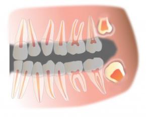
The norm for a person is the presence of teeth in the amount of 28–32 units. By the age of 25, complete formation of the dentition usually occurs. The teeth are located on both jaws, according to which the upper and lower dentition are distinguished. The structure of the jaw of a person, teeth (their typical classification) are as follows. Each row contains 14–16 teeth. The rows are symmetrical and they are conditionally divided into the left and right sectors. Teeth are designated by serial numbers - two-digit numbers. The first digit is the sector of the upper or
lower jaw, from 1 to 4.
During the closing of the jaws, the front teeth overlap the lower ones by 1/3 of the tooth crowns, and this ratio of dentition to each other is called a bite. With incorrect closing of the teeth, malocclusion is observed, which leads to a violation of the chewing function, as well as to an aesthetic defect.
So-called wisdom teeth may be absent and, in principle, may not appear in the oral cavity. Today, there is an opinion that this is a normal situation and the presence of these teeth is no longer necessary. Although this version causes a huge amount of controversy.
The teeth are not able to recover. Their change occurs once during a person’s life: first, the child has milk teeth, then at the age of 6–8 years they change to permanent. Usually, by the age of 11, there is a complete replacement of primary teeth with permanent ones.
The structure of the tooth. Anatomy
The anatomical structure of the human tooth suggests that it conditionally consists of three parts: the crown of the tooth, neck and root.
A tooth crown is a part of it that rises above the gum. The crown is covered with enamel - a strong fabric that protects the tooth from the damaging effects of bacteria and acids.
There are several types of surfaces of the dental crown :
- Occlusion - the surface at the junction with a pair of teeth on the opposite jaw.
- Facial (vestibular) - the surface of the tooth from the side of the cheek or lip.
- Lingual (lingual) - the inner surface of the tooth facing the inside of the oral cavity, i.e. the surface with which the tongue comes into contact when pronouncing sounds.
- Contact (approximate) - the surface of the dental crown facing the teeth in the neighborhood.
The neck is the part of the tooth located between the crown and the root, connecting them, covered with the edges of the gums and covered with cement. The neck has a narrowed shape.
A root is a part of a tooth, with the help of which it is attached in a tooth hole. Depending on the classification type of tooth, the root may have from one to several processes. This issue will be considered in more detail below.
Histological structure
The histology of each tooth is exactly the same, however, each of them has a different shape in accordance with the function performed by it. The figure demonstrates very clearly the layered structure of human teeth. The photo shows all the dental tissues, as well as the location of the blood and lymph vessels.
The tooth is enameled. This is a strong fabric, consisting of 95% of mineral salts, such as magnesium, zinc, strontium, copper, iron, fluorine. The remaining 5% is organic matter - proteins, lipids, carbohydrates. In addition, the enamel contains liquid that is involved in physiological processes.
Enamel, in turn, also has an outer shell - a cuticle, which covers the chewing surface of the tooth, however, over time it tends to thin out and wear out.
The basis of the tooth is dentin - bone tissue - a combination of minerals, strong, surrounding the cavity of the entire tooth and root canal. Dentin tissue includes a huge number of microscopic channels through which metabolic processes occur in the teeth. The channels transmit nerve impulses. For reference, 1 sq. mm dentin includes up to 75,000 tubules.
Pulp. Periodontium. Root structure
The inner cavity of the tooth is formed by the pulp - soft tissue, loose in structure, penetrated through by blood and lymph vessels, as well as by nerve endings.
The structure of the roots of human teeth looks like this. The root of the tooth is located in the bone tissue of the jaw, in a special hole - the alveolus. The root, like the crown of the tooth, consists of mineralized tissue - dentin, which is covered on the outside with cement - a fabric that is less durable compared to enamel. The tooth root ends with the apex, through the hole in which the blood vessels that feed the tooth pass. The number of roots in a tooth varies in accordance with its functional purpose, from one root in the incisors to 4-5 roots in the chewing teeth.
Periodontium is a connective tissue that fills the gap between the tooth root and the hole in the jaw in which it is located. Fibers of tissue are woven into the root cement on the one hand, and into the bone tissue of the jaw on the other, so that the tooth is firmly attached. In addition, through the periodontal tissue, blood vessel nutrients enter the tooth tissue.
Types of teeth. Incisors
Human teeth are divided into four main groups:
- incisors (central and lateral);
- fangs
- premolars (small chewing / molars);
- molars (large chewing / molars).
The jaw of a person has a symmetrical structure and includes the same number of teeth from each group. However, there are some anatomical features in such a matter as the structure of human teeth in the upper jaw and teeth of the lower row. Let's consider them in more detail.
The front teeth are called incisors. A person has 8 such teeth - 4 on top and 4 on the bottom. Cutters are designed to bite food, divide it into pieces. A special structure of the front teeth of a person is that the incisors have a flat crown, in the form of a chisel, with fairly sharp edges. Three tubercles protrude anatomically on the slices, which tend to wear off during life. On the upper jaw there are two central incisors - the largest of all representatives of their group. Lateral incisors are similar in structure to central ones, however, they are smaller. Interestingly, the cutting edge of the lateral incisor also has three tubercles, and often takes on a convex shape due to the development of the central (middle) tubercle. The root of the incisor is single, flat and takes the form of a cone. A characteristic feature of the tooth is that from the side of the tooth cavity there are three tops of the pulp, which correspond to the tubercles of the cutting edge.
The structure of the upper teeth of a person is slightly different from the anatomy of the teeth of the lower row, i.e., everything is exactly the opposite on the lower jaw. The central incisors are smaller in comparison with the lateral, have a thin root, shorter than that of the lateral incisors. The front surface of the tooth is slightly convex, but the lingual is concave.
The crown of the lateral incisor is very narrow and curved to the lips. The cutting edge of the tooth has two angles - the central, sharper, and the lateral - more blunt. The root is characterized by longitudinal grooves.
Fangs. Chewing teeth
Fangs are designed to separate food into smaller pieces. The tooth anatomy is such that a furrow runs on the back (lingual) side of the crown, which disproportionately divides the crown into two parts. The cutting edge of the tooth has one developed pronounced tubercle, which makes the shape of the crown conical, often similar to the fangs of predatory animals.
The canine of the lower jaw has a narrower shape, the edges of the crown converge in the medial tubercle. The root of the tooth is flat, the longest in comparison with the roots of all other teeth and tilted inward. A person has two fangs on each jaw, one on each side.
The fangs together with the lateral incisors form an arc, in the corner of which the transition from cutting to masticatory begins.
Let us consider more carefully the structure of the molar of a person, first - small chewing, then large chewing. The main purpose of chewing teeth is a thorough mechanical processing of food. This function is performed by premolars and molars.
Premolars
The first premolar (indicated by the number 4 in the dental formula) differs from the canine and incisors in its prismatic shape, the crown has convex surfaces. The chewing surface is characterized by the presence of two tubercles - buccal and lingual; grooves pass between the tubercles. The buccal tubercle is much larger than the lingual in size. The root of the first premolar is still flat, but it already has a bifurcation into the buccal and lingual parts.
The second premolar is similar in shape to the first, however, its buccal surface is much larger, and the root has a conical shape, compressed in the anteroposterior direction.
The chewing surface of the first lower premolar is beveled towards the tongue. The tooth crown is round, the root is single, flat, with grooves on the front surface.
The second premolar is larger than the first due to the fact that both tubercles are equally developed and symmetrical, and the depressions in the enamel (fissure) between them take the shape of a horseshoe. The root of the tooth is similar to the root of the first premolar.
In the dentition of a person there are 8 premolars, 4 on each side (on the upper and lower jaws). Consider the anatomical features and in general the structure of the human teeth of the upper jaw (large chewing teeth) and their differences from the structure of the teeth of the lower jaw.
Molars
The first molar of the upper jaw is the largest tooth. It is called a large
molar. The crown resembles a rectangle, and the chewing surface is a rhombus shape with four tubercles, between which the fissure is H-shaped. This tooth is characterized by three roots: one straight - the most powerful, and two buccal - flat, which are deflected in the anteroposterior direction. These teeth, when the jaws are closed, abut against each other and are a kind of "limiter", and therefore undergo tremendous stresses during the life of a person.
The second molar is smaller than the first. The crown has a cubic shape with an X-shaped fissure between the tubercles. The roots of the tooth are similar to the roots of the first molar.
The structure of human teeth (the location scheme of the molars and their number) fully coincides with the location of the premolars described above.
The first molar of the lower jaw has five tubercles for chewing food - three buccal and two lingual with an F-shaped fissure between them. The tooth has two roots - the back with one channel and the front with two. In addition, the anterior root is longer than the posterior.
The second molar of the lower jaw is similar to the first molar. The number of molars in a person coincides with the number of premolars.
The structure of the tooth of human wisdom. Baby teeth
The third molar is popularly called the "wisdom tooth", and in the dentition of a person there are only 4 such teeth, 2 on each jaw. In the lower jaw, the third molar can have many options for the development of tubercles. Often there are five. But in general, the anatomical structure of the “wisdom tooth” of a person is similar to the structure of the second molar, however, the root most often resembles a short and very powerful trunk.
As noted earlier, a person first has milk teeth. They usually grow by 2.5–3 years. The number of temporary teeth is 20. The anatomical and histological structure of the human milk tooth is similar to the structure of the permanent, but there are some differences:
- The size of the crown of primary teeth is much smaller in comparison with the permanent ones.
- The enamel of milk teeth is thinner, and the dentin composition has a lower degree of mineralization compared to molars, therefore, children often have caries.
- The volume of the pulp and root canal of a milk tooth is much larger in comparison with the volumes of a permanent tooth, which is why it is more susceptible to various inflammatory processes.
- The tubercles on the chewing and cutting surfaces are weakly expressed.
- The incisors of milk teeth are more convex.
- The roots are bent towards the lips, they are not so long and strong in comparison with the roots of permanent teeth. In this regard, the change of teeth in childhood is almost a painless process.
In conclusion, I want to note that, of course, the structure of a person’s teeth, the scheme of their location in the jaw, closure (occlusion) have individual characteristics characteristic of each particular person. However, the dentition of any person is involved in the implementation of vital functions of the body throughout life, in accordance with this, over time, the structure of the teeth and their structure change. It must be remembered that most pathological processes in dentistry develop in childhood, so it is important to monitor the condition of the teeth from the first years of life. This will help to avoid dental problems at a conscious age.
Despite the apparent simplicity, the teeth are a very complex and quite fragile system, with a multi-layered histological structure, each of the layers has an individual purpose and has certain properties. And the fact that the change of teeth occurs only once during life makes the structure of the jaw of a person (teeth, their number) different from the anatomy of the jaw of representatives of the fauna.