The human heart is one of the most perfect organs of the body, which performs important functions in the body. It has incredible power, distilling a large amount of blood through the arteries and small blood vessels per day. The heart is the motor in the human body. And it’s hard to disagree. But few know what the configuration of the heart is. Before considering this issue, you need to pay attention to what constitutes an organ, and what are its functions in the human body.
Description of the organ and its function
The heart is a hollow organ composed of muscles. With the help of rhythmic contractions, he provides the movement of blood through the vessels. The organ cavity is divided into several main parts. The left ventricle and atrium form the so-called arterial heart, and the right ventricle and atrium form the venous heart. The main function of the organ is to ensure continuous blood flow, therefore it distills the blood throughout the body, saturating the tissues and organs with oxygen and nutrients.
Heart shape
The size and configuration of the heart depends on the structure of the body, chest, respiratory activity, as well as the position of the body. The structural effects of heart disease also have an effect. The configuration also depends on the age, gender, health of a person. What are the parameters of the body:
- The length of an adult's organ can be from 10 to 15 cm, on average this figure does not exceed 12 cm.
- The width of the base can be from 8 to 11 cm, an average of 10 cm.
- The anteroposterior size averages 7 cm, but can be observed from 6 to 8.5 cm.
An important point of diagnosis is the sizing and determination of the configuration of the heart. They should be checked by all possible diagnostic methods. Thanks to this, specialists have the opportunity to make the correct diagnosis for various diseases of this organ.
Normal heart configuration
The human heart is presented in the form of a cone, slightly compressed. The top of the organ is rounded and facing down, forward and to the left. In humans, the heart is asymmetrical: 2/3 of its parts are located to the left of the middle of the body, the rest is located to the right of the middle plane. Its other placement is considered a deviation from the norm.
The septum, which separates the ventricles and atria, is located in a healthy person between the sagittal and frontal planes. The right atrium and ventricle, pulmonary artery and aortic arch, as well as part of the left ventricle are located on the frontal plane. Behind the organ is another part of the left ventricle and the left atrium, as well as part of the right ventricle. According to the physique of a person and the shape of his chest, conclusions are drawn about what size he has and whether his heart is normal.
Heart shape
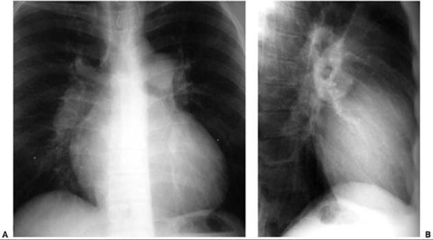
In the process of diagnosis, the location of the right and left contour of the organ is determined. Where should they be visible? The right contour should be observed in the upper part of the chest from the first intercostal space to the third third rib, and the left contour is represented by the first and second intercostal space, the left atrium and further down the narrow strip of the left ventricle.
After the configuration of the heart is determined, the measurement of the length and diameter is carried out. What it is? The length is the distance from the top point of the left contour and the apex of the right cardiovascular corner. Usually it is 13 cm in men and 12 cm in women. The diameter is measured by the distance from the most distant point of the right and left contours to the midline of the heart. In men, it is 11 cm, in women - 10. Then, between the diameter and the length, the angle of the organ is measured, which makes it possible to talk about its position:
- the middle position is indicated by a slope of 30 to 50 degrees,
- horizontal position - from 30 degrees or less,
- vertical position - from 60 degrees or more.
Determining the contours of the heart in a person makes it possible to draw conclusions about the causes that caused their change.
Changing the configuration of the heart
In pathology, there is a description of five changes in the shape and position of the heart. Let's consider in more detail each of them:
- The aortic configuration of the heart is observed in humans with severe left ventricular hypertrophy. This phenomenon is characterized by a shift of the lower part of the left contour outwards. The diameter and the length of the increase while the angle of inclination decreases. These changes are observed with malformations of the aortic valve, stenosis of the aortic orifice, hypertension, and cardiomyopathy.
- Spherical heart arises as a result of hypertrophy of the right ventricle. Changes cause a septal defect. There is a shift of the lower part of the right contour outwards. The diameter and angle of inclination increase, the length remains normal. Such changes can be congenital and observed with pericarditis, interventricular septal defect, narrowing of the pulmonary artery, and with a three-chambered heart. Also, such an organ is often found in athletes, as well as in children and adolescents.
- Mitral configuration of the heart is observed in people who have mitral stenosis. They have hypertrophy of the left atrium and right ventricle, as a result of which there is a shift in the lower part of the right contour outwards. At the same time, the angle of inclination and the diameter increase, the length remains normal. Such changes are also observed with defects of the mitral valve, a change in the myocardium, and a violation of the diastolic function of the myocardium.
- A bull heart is inherent in people who have a strong increase in all heart chambers. This occurs in the presence of heart defects and dilated cardiomyopathy.
- The trapezoidal configuration of the heart is observed when fluid accumulates in the pericardial cavity. In this case, there is a shift of the lower part of the left and right contours outwards. Also, the cavity may contain air in the presence of an abscess or decaying tumor. Such phenomena are often observed in various cardiovascular diseases. This may be obesity, pericarditis, myocarditis, cardiosclerosis and so on. In some cases, this configuration is observed in children with a high diaphragm. In this case, this is considered the norm.
Diameter change
The diameter is a term of two transverse dimensions of an organ (right, left). So, the configuration of the heart in a healthy person suggests its presence, as well as the size of this part equal to 11 to 13 cm. The right parameter is measured by the distance from the right border to the front midline. It should be normal 3 or 4 cm.
The size of the left is determined by the distance from the left border to the front midline. It should normally be 8 or 9 cm. An increase in the right size of the diameter occurs with pathology, which is accompanied by dilatation of the right atrium and ventricle. Pericarditis also leads to the development of pathology. A change in the left cross-sectional size occurs with disorders that are accompanied by dilatation of the left ventricle.
Vascular bundle change
The cardiac contours, which are determined in the second intercostal space from all sides, correspond to the size of the vascular bundle. In a healthy person, its right side runs along the right border of the sternum. At the end of the vascular bundle, an aorta is formed. The left border runs along the left edge of the sternum. Here at the end of the vascular bundle is formed by the pulmonary artery. The width of the site is 5 or 6 cm. An increase in its size occurs with the development of atherosclerosis and aortic aneurysm, while the configuration of the heart also changes.
Other causes of changes in the vascular bundle are associated with diseases, which are accompanied by the appearance of additional tissue. This may be, for example, goiter, enlarged lymph nodes, the presence of primary tumors or metastases. The expansion of the vascular bundle appears with atherosclerosis of the aorta, aortic aneurysm, expansion of the pulmonary artery, and an increase in blood pressure.
X-ray diagnosis
The shape of such an organ as the heart is of great importance. Therefore, its X-ray diagnosis is often carried out. The most common heart diseases are defects, myocardial pathology. Such violations lead to the fact that the configuration of the heart on the roentgenogram changes. This helps to make an accurate diagnosis, which is the main thing in the appointment of appropriate treatment. Diagnostics makes it possible to solve questions on the assessment of myocardial perfusion in heart disease.
Studying the configuration of the heart is an urgent problem today. Modern diagnostic methods make it possible to detect even minor changes in the size and location of the organ, which can lead to various complications. When conducting an x-ray, the severity of the arcs is analyzed, which form the edges of the cardiovascular bundle, as well as their delimitation along the left contour. If the configuration of the heart is normal, then, to diagnose disorders in the body, other organs are examined.
In conclusion
Thus, a change in the boundaries of an organ is caused by the presence of various cardiac and vascular diseases. This includes, for example, tricuspid valve insufficiency, aortic heart defects, heart failure, pneumothorax, myogenic dilatation, etc. Also, a change in configuration may be a consequence of the asthenic type of physique. Modern diagnostic methods allow you to make an accurate diagnosis in order to prescribe an effective treatment for various heart diseases.