As you know, during pregnancy, the future man undergoes fundamental transformations - from a tiny fertilized egg to a complex organism, capable of independent life outside the mother’s womb. As it grows, the space in the uterus becomes smaller. The child can no longer move freely inside her and occupies a certain position, more or less constant (as a rule, after the 32nd week it no longer changes).
To describe the placement of the fetus in the uterus in late pregnancy and immediately before childbirth, specialists use three characteristics. This is the type of position, position and presentation of the fetus. It directly depends on how the birth will take place - either naturally or with the help of a cesarean section, and also what difficulties may arise during this process. These characteristics will be discussed in the article.
Item type
The following types of fetal position are distinguished: front and rear. With the front, the fetal back is turned anteriorly, with the rear, respectively, posteriorly.
What is presentation
The term "presentation of the fetus" is used to describe how the child is located in relation to the entrance to the small pelvis. Buttocks or the baby’s head can be turned to it. Head previa is the most common, it occurs in almost 97% of cases. This is the most favorable, correct position of the fetus for natural childbirth.
Head presentation: types, characteristics
There are several types of head presentation, and not all of them are equally good for self-delivery. The most natural is the occipital, in which the fetal head is cut, respectively, by the back of the head, with a front view of the position, that is, one in which both the back and back of the fetus are facing forward. Some of the species, namely, the anterior-head, frontal and facial, are relative indications for cesarean section. These are the so-called extensor presentation.
Their causes may include shortening of the umbilical cord, clinically and anatomically narrow maternal pelvis, decreased uterine tone, small or too large fetus size, stiffness of its atlanto-occipital joint, etc.
Extensor type of labor mechanism
The extensor types of presentation, in which the fetal head is moved to one degree or another from the chin, are diagnosed with an internal vaginal examination of the mother. All of them pose a certain danger to the mother and the fetus, leading to prolonged childbirth and complications. There are three types of extensor presentation, depending on the degree of extension of the head: anterolateral, frontal and facial.
Facial presentation
The case, opposite in all characteristics to the anterior occipital presentation, is the so-called facial presentation, in which the fetus comes out with the chin forward and an extreme, maximum degree of extension of the head is noted. The nape may literally lie on the child’s shoulder girdle. Facial presentation is rare (0.5%). Most often, this type of presentation occurs directly during childbirth (secondary), it is extremely rare that it is established during pregnancy (primary). The head in this case is cut through by the so-called face line, conditionally connecting the center of the forehead with the chin, and having reached the pelvic floor, it is bent forward by the chin.
Despite the complexity, 95% of such births end on their own. Five percent of cases require emergency care. After childbirth, facial presentation for 4-5 days in the newborn retains swelling of the face and characteristic extension of the head.
Frontal presentation
This type of presentation is quite rare, in about 0.1% of cases. It is extremely traumatic, childbirth is characterized by a protracted course (up to a day in primiparas) and end in fetal death, according to various sources, in 25-50% of cases. According to statistics, only in a little more than half of the cases (approximately 54%) a natural birth is possible without surgery. The severity of their course is due to the fact that it is in the frontal presentation that the fetus must pass through the pelvis with the largest plane. For a woman in childbirth, the slow advancement of the fetus through the birth canal is fraught with ruptures of the perineum and uterus, the appearance of fistulas and other complications.
The established stable frontal presentation of the fetus is currently considered to be one hundred percent indication for cesarean section, which, in turn, is possible provided that the fetus has not yet had time to fix in this position at the entrance to the pelvis. Since most often this position of the fetus is unstable, and is usually transitional from the anteroposterior to the facial, during childbirth it can spontaneously go both to the occipital (rarely) and facial, therefore, the choice of expectant management tactics of childbirth makes sense. However, it is extremely important not to miss the time for cesarean section.
Forehead Presentation
With this presentation, the degree of extension of the head is the smallest possible (the chin is slightly moved away from the chest). Primary anterior-head presentation is extremely rare, its cause is the presence of a thyroid tumor in the child. More often it occurs during childbirth.
It can be determined by the palpable large and small fontanel, while with the occipital presentation during examination, only a small fontanel is available. The head cuts through the area of the large fontanel, that is, a circle that corresponds to its direct size. A birth tumor in a child is usually also located in this area.
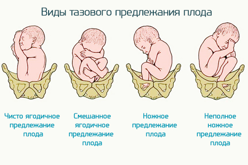
Pelvic presentation
Pelvic is a type of presentation in which the fetus is located at the pelvic end to the entrance to the small pelvis of the woman in labor. The frequency of this pathology, according to various sources, may be 3-5%. Childbirth in this position is fraught with complications for both the mother and the child.
There are three main types of it:
- Gluteal - the fetus is located buttocks down, legs are bent, knees are pressed to the stomach (up to 70% of cases).
- Leg (may be complete or incomplete) - one or both legs are unbent and located near the exit from the uterus.
- Mixed - hips and knees bent (up to 10% of cases).
Pelvic presentation has no external signs by which a pregnant woman could determine it. Only the ultrasound study after the 32nd week can give an accurate picture. If the pelvic presentation has not been determined in advance, during a vaginal examination during childbirth, the doctor can determine it, depending on the type, by the palpable parts - the coccyx, buttocks, and the feet of the fetus.
During delivery, a cesarean section is most often recommended. The decision to choose an operative method or natural birth is made based on several indicators: the age of the expectant mother, the presence of certain diseases, the course of pregnancy, the size of the pelvis, the weight of the fetus and the type of presentation, and the condition of the fetus. When a boy is pregnant, preference is given to cesarean section, since the likelihood of complications in this case is higher. Most likely, such a decision will be made in the case of leg presentation, as well as if the fetus weighs up to 2500 or more than 3500 g.
If complications arise during the natural birth in the pelvic presentation, such as placental abruption, fetal hypoxia, loss of body parts or the umbilical cord, a decision is made on emergency cesarean section. This is true for a situation where there is a weak labor activity and childbirth, respectively, is delayed.
What is the position of the fetus
There are such types of fetal position: longitudinal, transverse and oblique. In the first case, the axis of the fetal body is located along the longitudinal axis of the woman’s uterus. In the second, respectively, across it. The oblique position is intermediate between the longitudinal and transverse, while the fetus is located diagonally. The position of the fetus is the longitudinal head - normal, physiological. It is most favorable for childbirth. The transverse, as well as oblique, are classified as incorrect fetal position (photo can be seen later in the article).
Oblique and lateral position of the fetus
They are unfavorable for natural childbirth. In the transverse and oblique position of the fetus, the underlying part is not determined. Such situations are possible in about 0.2-0.4% of women in labor. The reason for them, as a rule, are health problems in women (uterine tumors), uterine overstrain due to multiple births, as well as entanglement of the umbilical cord in the fetus or its large size. A short umbilical cord is another possible reason for adopting this position.
With the transverse position of the fetus, pregnancy can proceed without complications, however, there is a risk of premature birth. Complications are also possible: water leakage, uterine rupture, loss of parts of the fetus.
The optimal solution for transverse and oblique position of the fetus will be operative delivery using cesarean section. A woman in labor is hospitalized two to three weeks before the expected date of delivery in preparation for surgery.
Ways to Correct Provisions
With a pelvic presentation, an oblique and transverse position of the fetus, it is possible to perform pregnant special gymnastics in order to correct them. Exercises may be permitted by the doctor in the absence of contraindications, such as:
- Placenta previa.
- Multiple pregnancy.
- Hypertonicity of the uterus.
- Myoma.
- Scar on the uterus.
- The presence of serious chronic diseases in the woman in childbirth.
- Low water or high water.
- Bloody issues
- Gestosis and others.
Exercise should be combined with deep breathing. The complex may look like this:
- Lying on your back, raise the pelvis above the shoulder level by 30-40 cm and hold it in this position for up to 10 minutes (the so-called "Half Bridge").
- Standing on all fours, tilt your head. On inhaling, round your back, while exhaling, bend in the lower back, raising your head up (this exercise is often called the "Cat").
- Rest your knees and elbows on the floor so that your pelvis is above your head. Hold in this position for up to 20 minutes.
- Turn over from side to side, lingering on each for 10 minutes.
With the oblique position of the fetus, it is recommended to lie down more often on the side where its back is turned.
It should be remembered that exercises to correct the position of the fetus can be done only on the recommendation and with the permission of the doctor. He can advise other exercises. Due to corrective gymnastics, the fetus can take the correct position for 7-10 days. Otherwise, it is considered ineffective.
External obstetric turn to change the position of the child (according to B. A. Arkhangelsky)
In a hospital for a period of 37-38 weeks, it is possible to perform the so-called external obstetric rotation of the fetus, which is performed by external methods, through the abdominal wall, without penetrating the vagina and uterus. In this case, the obstetrician has one hand on the head, the other on the pelvic end of the fetus and turns the buttocks towards the back, and the head - towards the abdomen of the child. Currently, this procedure is practically not used. This is due to its low efficiency, since the fetus can take its former position if its causes have not been eliminated. In addition, there is a likelihood of serious complications: the development of fetal hypoxia, placental abruption. In rare cases, even a uterine rupture is possible. Therefore, the rotation of the fetus can be recommended only with normal fetal mobility and the normal amount of water, the normal size of the pelvis and the absence of pathologies in the pregnant woman and the child.
Manipulation is carried out under the control of an ultrasound machine using injections that relax the muscles of the uterus (ß-adrenergic agonists).
Leg rotations, which were widely used earlier during childbirth, are now practically not used, since they can be of great danger to the mother and fetus. Their use is possible with multiple pregnancy, in the event that one of the fruits takes the wrong position.
After the transition of the fetal position to the head, right, pregnant woman, it is recommended to wear a special bandage with rollers to fix the child. It is usually worn until birth. If the methods described above for correcting the position of the fetus did not work, two or three weeks before the expected date of birth, the woman is hospitalized and the question of choosing a natural or operative method of childbirth is decided.
Situation with multiple pregnancy
When there are several babies in the uterus, it may be difficult for them to take the correct position due to lack of space. In pregnancy, twins are possible when both fetuses take the correct position, or one of them is presented with the pelvic end to the exit from the uterus. Much less common are cases when they are in different positions (longitudinal and transverse), or the location of both fetuses is perpendicular to the axis of the uterus.
In the normal course of childbirth, after the birth of the first of the babies, there is a pause in labor activity lasting from 15 to 60 minutes, and then the uterus adapts to a reduced size, and childbirth resumes. After the appearance of the second child, both afterbirths are born.
The following complications are possible in childbirth during a multiple pregnancy: the first fetus waters leave before labor begins, its weakness, accompanied by delayed childbirth, the so-called twin cohesion, etc. If the position of one or both fruits is incorrect, the situation is even more complicated. The decision on the method of delivery must be made by the doctor, since in many cases natural births are dangerous for both the mother and the babies.
Finally
As can be understood from the above, the position of the fetus, its position and presentation are the main characteristics that are taken into account by doctors when choosing a method of childbirth. It should be understood that in certain situations, natural childbirth is fraught with great complications. Therefore, if a specialist decides to have a cesarean section, you must trust him. This will protect both the mother and the child from serious health problems in the future.