Many confuse the concepts of veins and arteries. Let's look at how these two elements of the human circulatory system differ from each other, before moving on to an overview of its specific part.
Heart
The central organ of the human vascular system is the heart, to which the tubes of various sizes and diameters, the so-called blood vessels, are closed. By rhythmically contracting, it pumps the blood inside the body. Arteries are called vessels that carry blood from the heart to peripheral organs, while veins deliver blood back to the heart. This is the main difference. Venous and arterial bleeding are distinguished by characteristic signs: during the first, blood flows in a stream, and in the second it gushes.
Arteries and veins
Several cardinal differences of arteries and veins are distinguished:
- Arteries carry blood to organs from the heart, veins in the opposite direction. in the first case, oxygen is transported through the vessels, and in the second, carbon dioxide.
- In arteries, the walls are thicker and more elastic than in veins. Blood in them moves under pressure. In the veins, the flow is much calmer.
- The veins are twice as large as the arteries, and their location is more superficial.
- In the medical field, veins, rather than arteries, are used to collect test materials.
This article will examine the femoral vein.
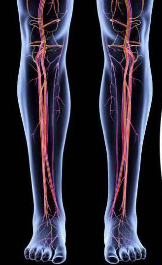
Venous network
To conduct the correct diagnosis of the disease and make the correct diagnosis in the field of venous diseases, you need to clearly understand the system of blood vessels of the lower extremities. Distinguishes a deep and superficial network of veins. Deep consists of paired vessels passing near the arteries on the fingers, foot and lower leg. The tibial veins converge in the femoral-popliteal canal and create an unpaired popliteal vein passing into the femoral vein. Before the transition to the iliac, up to 8 peripheral vessels join it. In addition to them, adds a deep vein that carries blood cells from the back of the thigh.
The superficial circulatory network is located directly under the skin. It consists respectively of a large and small saphenous vein.
Veins hips
It is extremely important for a vascular surgeon to know the detailed structure of the circulatory system. If the vessel consists of several trunks, it is difficult to find a deep femoral vein. Surgeons conditionally divide it into a surface, located more deeply, and general, which is closer to the confluence of a deep vein.
Deep vein is the farthest of all major tributaries. It connects to the femoral vessel just below the inguinal region. Further, smaller tributaries enter it. In addition, two additional, called the paraarterial venous bed, fall into the lower part of the mouth of the deep vein.
Common vein
The total femoral vein includes the large saphenous, medial and lateral veins surrounding the thigh. Each has its own location and meaning. The medial is closer than the lateral. She joins in the area of large subcutaneous and higher.
In the vein of the thigh, there are usually up to 5 valves preventing the blood from moving in the opposite direction. The distance between them often reaches 7 cm. In this case, the clearance is often not more than 12 mm. Sometimes she has two trunks that connect at the bottom of the sciatic tubercle. The deep femoral vein is located on the outside of the femur, which its proximal section crosses, flowing into the main.
The veins accompanying the femoral artery are located in the lower and middle parts of the thigh, on the external or internal side of the artery and are connected to it in several places. Such sites are called anastomoses. Depending on how the valves are located in the vessels accompanying the femoral artery, blood can flow in them in different directions.
A large saphenous vein happens with two or more trunks. True doubling is the case when it enters the femoral with different mouths. But much more often they connect in the upper thigh. The anatomy of the femoral vein is considered by us.
Pathology
The most common diseases of the veins of the thigh are thrombosis and venous expansion. And if the latter disease is ubiquitous and in most cases does not threaten life, although it is rather unpleasant, then thrombosis is another matter. It is worth talking about it separately.
Thrombosis
Femoral vein thrombosis is of two types: superficial and deep. Such a deep vein disease is the formation of blood clots that partially or completely clog a vessel. Most often this happens in the lower extremities. More specifically, in the veins of the thigh. 20% of the population of our country suffer from this disease. The bulk of the disease occurs in men, quite rarely in women (mostly suffering from varicose veins). Without proper treatment, deep vein thrombosis can be fatal due to pulmonary embolism.
Signs of superficial femoral vein thrombosis are:
- Swelling and pain in the legs, starting from the groin and below.
- Cyanosis of the skin on the legs.
- The so-called petechial rash in the form of small red dots.
- An increase in body temperature as a result of phlebitis - inflammation of the walls of the vessel.
With deep vein thrombosis, two stages are distinguished: white and blue phlegmasia. At the initial stage, due to a violation of blood circulation, the skin of the leg becomes pale, cold to the touch, with severe pain.
Blue phlegmasia is a sign of overflow of venous vessels with blood. With it, the skin may darken, and on its surface there may be swelling, which contains hemorrhagic fluid. With these symptoms, thrombosis runs the risk of flowing into acute gangrene.
Background of Deep Vein Thrombosis
Most often, deep vein thrombosis occurs when a vessel is compressed for a long time by a tumor or bone fragment during a fracture. Another reason for the formation of cork is a violation of blood circulation in certain diseases. Poorly circulating blood leads to stagnation and, accordingly, blood clots. The key causes of vein blockage are as follows:
- Drop in blood circulation in the vessels.
- Increased coagulation time.
- Damage to the walls of blood vessels.
- A long stay in a stationary state, for example, with a serious illness.
A negative effect on the condition of the veins is exerted by some professional activity. Salesmen, cashiers, pilots, international drivers have a hard time. They are forced to stand or sit in one position for a long time. Therefore, they are at risk. Frequently recurring diseases that lead to dehydration, such as acute intestinal infections, accompanied by diarrhea and vomiting, chronic diseases of the intestines and pancreas. It also occurs against the background of excessive medication with a diuretic effect. Pathologies that cause an imbalance of fats and proteins, including diabetes, atherosclerosis, and cancer, are dangerous. Bad habits lead to an increased likelihood of platelet sticking: smoking, alcohol abuse.
What is femoral vein catheterization needed for? About it below.
Diagnosis and treatment
It is not worth talking about the importance of timely diagnosis and medication or other intervention in DVT. To make an accurate diagnosis, it is necessary to do an ultrasound or dopplerography of the femoral vein. Such diagnostics will help determine the exact location of the thrombus and the degree of its fixation to the vessel wall. In other words, to understand whether it can come off and clog the vessel, as well as cause pulmonary thromboembolism or not. Also, in the detection of DVT, the phlebography method is used - an x-ray with a contrast medium. However, the most accurate method to date is angiography. On the eve of the procedure, you must comply with strict bed rest. Sometimes a femoral vein puncture is performed.
Treatment for DVT depends on the cause of the disease and the individual characteristics of the patient. If the vessel is not completely blocked and a thrombus detachment is unlikely, conservative therapy is indicated. It is necessary to restore the patency of the veins, prevent a violation of the integrity of the thrombus and avoid embolism of blood vessels. To achieve the above goals, they use special medications, ointments, as well as compression therapy, for example, they recommend wearing special compression stockings.
If the patient is in a satisfactory condition, but medication is contraindicated, then surgical methods of treatment of deep thrombosis are used. The operation is carried out on the latest equipment and is high-tech. Thrombectomy is prescribed when the risk of separation of the thrombus and clogging of the main vessels is not excluded. Such a plug is removed through a small incision by introducing a special catheter. During the operation, the "clogged" vessel is completely cleaned, but relapse is not excluded.
To avoid thrombosis, you must adhere to some rules and completely redefine your lifestyle. It is recommended to abandon bad habits, eat right, lead a physically active lifestyle, try to avoid injuries of the lower extremities, etc. We examined the femoral arteries and veins. Now you know how they differ and what they are.