One of the necessary conditions for the onset of pregnancy is the normal patency of the fallopian (or uterine) tubes in a woman. After all, it is through these channels that the fertilized egg penetrates the uterus. If patency is impaired, then a woman is diagnosed with infertility. In the event of partial obstruction, a life-threatening condition may occur - an ectopic pregnancy. To protect a woman from such problems and assess her chances of conceiving a child, the doctor prescribes an ultrasound of the patency of the fallopian tubes.
Brief characteristics of the survey
To assess the patency of the fallopian tubes, the patient is prescribed an ultrasound of the patency of the fallopian tubes, or, in the language of doctors, hysterosalpingoscopy (GHA). This is a special diagnostic study, which, with the help of a contrast agent, allows you to explore the female genital area.
Unfortunately, conventional ultrasound is not able to fully provide information about the passage of pipes. That is why doctors resort to a special technique that detects unpleasant pathologies.
Ultrasound (GHA) of the fallopian tubes can be performed in two ways:
- transvaginally (by insertion into the vagina);
- when using an external sensor.
This examination method is completely safe and very effective.
Indications for the study
Any alarming signs, incomprehensible pains in the lower abdomen, the appearance of deviations in the cycle are serious reasons to go to the gynecologist for a consultation. If necessary, the doctor will prescribe a woman with an ultrasound of the fallopian tubes for patency. However, the doctor gives this direction only after a gynecological examination.
Ultrasound (GHA) of the fallopian tubes is recommended for women who have the following pathologies:
- irregular menstrual cycle (irregularity or lack of menstruation);
- infertility;
- transmitted infections that are transmitted sexually;
- persistent pain in the lower abdomen;
- inflammation of the appendages.
In such conditions, an examination may be prescribed after treatment. This allows you to determine the effectiveness of the prescribed therapy and assess the condition of the woman. Ultrasound patency of the fallopian tubes can be performed repeatedly. After all, such a study is painless and does not harm women's health.
Dates
To get the most reliable clinical picture, you need to choose the right days. This is very important when an ultrasound scan is performed. Gynecology (which days are considered the most suitable, we will describe below) is a special branch of medicine that requires responsibility on the part of the doctor, because the health of not only a woman, but also her future offspring depends on his actions.
Doctors recommend conducting a study taking into account such terms:
- The period from the 6th day of the cycle to the 21st.
- Some gynecologists advise an ultrasound scan before ovulation, from the 7th to the 12th day of the cycle.
Why are these terms considered the most optimal? Doctors say that in these periods the cervix is maximally expanded. The endometrium after menstruation has a minimal thickness. These features allow the most accurate research.
Study preparation
Ultrasound of the fallopian tubes for patency is a great chance to identify pathology at an early stage, timely start adequate therapy and completely get rid of the disease.
However, in order to correctly diagnose the condition of a woman, it is necessary not only to choose the appropriate dates for the procedure. It is equally important to properly prepare for the study. The gynecologist will tell you about all the necessary activities.
The preparatory stage for the survey usually includes the following activities:
- Putting a smear on the microflora of the vagina. Such an analysis is valid for 21-45 days. Be sure to check with your doctor for the duration of the smear.
- A blood test for such indicators: F-50 (for HIV), RW (for syphilis), hepatitis B, C. The results of these studies are considered valid for 6 months.
- Compliance with a special diet 3 days before the examination. During the event, it is necessary to exclude the presence of gases in the intestine. That is why it is important to adhere to a diet for 3 days, aimed at reducing fermentation in the digestive tract. Refuse flour, sweet, sour-milk. Do not eat vegetables, fruits, sodas, and legumes.
- Ensuring vaginal cleanliness. A week before the ultrasound, it is recommended to abandon the use of vaginal sprays, suppositories, tablets. Exclude douching.
- Acceptance of an antispasmodic. About 20 minutes before the start of the procedure, the patient will be advised to use an antispasmodic drug (Spazmalgon, No-Shpa). Such a medication will provide relaxation of smooth muscles and prevent reflex contractions of the uterus. Sometimes such drugs are administered to a woman before the injection procedure.
Survey
The procedure consists of several stages:
- Diagnosis of fallopian tubes begins with preliminary ultrasound monitoring of the condition of the pelvic organs. Such a study is carried out in order to exclude a woman's pregnancy and the presence of inflammation.
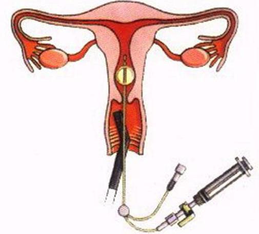
- The patient is comfortable in the gynecological chair. A disposable catheter is inserted intravaginally into the cervix. Through it, a contrast agent enters the organ cavity, preheated to a comfortable temperature (37 degrees). The solution fills the uterus and rises through the fallopian tubes. Then, the contrast medium moves into the abdominal cavity.
- It is at the third stage that the analysis of pipe passability begins. Free fluid localized in the pelvis indicates satisfactory patency. Significantly worse if the substance does not penetrate the peritoneum. Fluid that does not leave the pipe limits clearly indicates obstruction of the channels. This study also allows you to determine the speed of movement of the contrast medium along the ducts.
Ultrasound using a contrast agent makes it possible to assess the condition of the uterus and its structure:
- the shape and contour of the organ;
- the presence of polyps and myomatous formations;
- relief and thickness of the endometrium;
- development of the organ and pathology in its structure.
The duration of the study averages 25-30 minutes.
Survey results
During the procedure, a special sensor detects the presence of fluid in the peritoneum. The doctor begins the examination with the uterus. It then determines the condition of the fallopian tubes. After that, if necessary, completes the examination by studying the ovaries.
An ultrasound of the patency of the fallopian tubes reveals:
- congenital malformations;
- myomas, polyps, endometriosis, fibromas;
- adhesive processes (at the same time clearly defines the localization of the pathology);
- pipe contours;
- the location of the fallopian canals.
Contraindications to the study
Ultrasound on the patency of the fallopian tubes is a fairly simple procedure. It does not imply invasive intervention and does not require complex preparation for the conduct. But even such a simple examination has a number of contraindications.
It is strictly forbidden to conduct an ultrasound scan:
- with uterine bleeding;
- gynecological diseases occurring in acute or chronic form;
- violations of the vaginal microflora;
- inflammatory diseases of the pelvic organs;
- pregnancy
- tumors (benign, malignant) located in the pelvis;
- infectious pathologies during exacerbation.
Tubal patency analysis: research cost
How much does this examination cost women? Of course, depending on the chosen clinic, the cost of ultrasound on the patency of the fallopian tubes will also vary. The average price of the procedure is 5395 rubles.
Research benefits
GHA has a number of advantages in comparison with other diagnostic methods for patency of the fallopian tubes.
The main advantages of ultrasound are:
- The procedure does not require hospitalization.
- The introduced liquid disunits the spliced areas, and the derivative is washed off.
- During the study, many pathologies of the uterus are simultaneously detected.
- The egg is promoted.
- The patient does not need anesthesia.
- The study is carried out quickly enough, and the results are visible immediately.
- There is no need for peritoneal punctures.
Having noticed the first alarming symptoms, do not delay the visit to the gynecologist. Be sure to seek the help of competent specialists who, if necessary, will recommend you an effective and safe ultrasound scan.