The fifth week of pregnancy is the period with which they most often come to be registered in gynecology. Usually the delay of menstruation at this time is at least 14 days, now it can no longer be attributed to chance. Usually by this time signs of toxicosis begin to show themselves brightly. This is not to say that these are especially pleasant moments, but such is the first trimester. It is an ultrasound scan at 5 weeks gestation that is the first documented confirmation of this special condition, which is why doctors often refer women to the first examination to resolve all doubts and make a diagnosis.
How much ultrasound is needed for this period
Usually, doctors are in no hurry to send expectant mothers for examination. The term is still very small, and there is no urgent need to do an ultrasound at the 5th week of pregnancy. However, sometimes very impatient mothers themselves register for an examination, which today can be taken at any clinic, especially on a paid basis. Most of all, this applies to the category of women who have been waiting for pregnancy for a long time and want to get confirmation that they will really become mothers. Most often, at such a short time, the doctor will send an ultrasound scan if the woman wants to terminate the pregnancy. Therefore, if nothing bothers you, there is no bleeding, severe pain, it is better to wait a bit, because the study at this time may not be very reliable.
What is the uterus at this time?
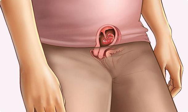
Externally, the expectant mother is no different from her friends, but miracles are already beginning to happen inside her. First of all, the uterus itself changes. Ultrasound confirms a pregnancy of 5 weeks only if it is carried out on good equipment. However, an experienced doctor can determine the increase in the uterus itself. This is a normal phenomenon, because inside it there is a fetal bladder larger than 1 cm. However, this increase is uneven: it is from the side where the embryo is attached to the uterus that protrudes. Ultrasound at 5 weeks of pregnancy is not very informative, so even an experienced specialist who works on good equipment can not say much besides the fact that you will be a mother. When the image is magnified several times, he will be able to identify the yolk sac and the embryo itself. Moreover, it was at this time that the baby's heart began to beat. In addition, at this time in the ovary, from which the egg came out, the corpus luteum is still noticeable, which is evidence of early pregnancy.
What is visible on the monitor
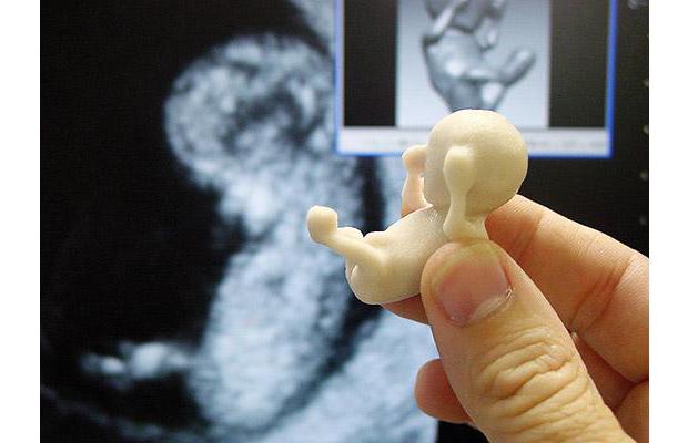
Not always a woman undergoes an ultrasound in a specialized clinic where there is special equipment for three-dimensional ultrasound. But on a regular device, no one except the specialist himself will be able to make out what he sees on the screen. Therefore, you will have to rely on the experience of the doctor. Ultrasound at 5 weeks of pregnancy allows you to see the embryo itself, which so far is very similar to a small cylinder. Its length is five millimeters, and its weight is only 3.5 grams. However, despite the fact that it is still very small, the most important processes occur inside it that determine all subsequent development. Now a neural tube is being formed, the respiratory system, liver and pancreas are being laid, cells are being formed, from which male and female germ cells will then develop. On the monitor, not only the tail and head are visible, but also the rudiments of the arms and legs, fingers and eye. With the help of a special sensor, the first heartbeat of a little man is heard.

First measurements
Ultrasound at 5 obstetric weeks of pregnancy allows you to make the first measurements and approximately determine whether the embryo is developing correctly. The coccyx-parietal size of the embryo is measured, based on this it will be possible to calculate the weight of the unborn baby during childbirth. Be sure the doctor will evaluate the position of the fetus in the uterus, the tone of the myometrium, the condition of the ovaries and corpus luteum. At the same time, the doctor on the monitor sees an enlarged uterus, resembling an egg in shape. Such an asymmetry appears due to implantation of a fertilized egg into the endometrium.
Embryo movements
It is amazing how much doctors can learn about the baby when the gestational age is 5 weeks. Ultrasound allows the doctor to conclude how active the embryo is. It is the frequency of his movements together with the heart rate that allows us to draw the first conclusions about his vitality and well-being. If the embryo is now without movement, then the doctor will raise the question of abortion. There are still several weeks to wait and compare the indicators at the next examination, but these are already alarming calls.
What is important for a future mother to know
Ultrasound at 4, 5 weeks of pregnancy is not always done, but at this time the woman usually already knows that she is pregnant. Therefore, it is extremely important to know about the features of the development of the embryo, as well as about the rules that a pregnant woman should adhere to. First of all, remember that just now the neural tube is completing its formation. Do you want your baby to be born healthy, calm and smiling? Then leave all the worries and troubles, and also drink any soothing herbal tea more often. We should not forget about folic acid preparations, which are extremely important for the proper development of the nervous system. In addition, this is exactly the period when you need to carefully monitor your health. The intake of most drugs during this period is prohibited, and the simplest cold threatens with serious pathologies of the fetus. For this reason, doctors are trying to dissuade you from taking the first ultrasound scan. 5 weeks of pregnancy is the time when you need to first think about the health of the baby, despite the fact that the harm of this procedure has not been proven, it is necessary to minimize all risks.
Opinion and reviews of expectant mothers
Usually this examination does not cause special emotions in women. Ultrasound at 5 obstetric week of pregnancy does not carry any information load. Everything that the doctor sees, while too rough, you can not find out either about the field of the future baby, or about how its development goes. However, if the doctor turns out to be a real specialist, as well as a kind and attentive person, he can tell you a lot about the features of the development of the embryo at this stage, the main milestones that you need to pay attention to. It is this examination that makes it possible to completely exclude an ectopic pregnancy and establish that the fetus develops exactly where it should be. But do not rush while doing a memory photo. An ultrasound of 5 weeks of pregnancy shows very schematically, so it is unlikely that you will be pleased with a striped background with obscure points in your family album. You will still have enough time and opportunities to capture the baby.
Is ultrasound harmful at this time?
In fact, no one will answer this question with full confidence. Too many components. There is still no reliable data that would confirm that this procedure is harmful to the embryo. However, speaking about whether an ultrasound scan is done at 5 weeks gestation, it should be noted that without serious reasons this procedure is not prescribed. This may be the patient’s complaints (pain, discomfort, bleeding) or a doctor’s suspicion. And, to eliminate the risk to the mother, the gynecologist can send for an ultrasound. Of course, in any paid clinic, they will do it even without a referral, especially if you say that you are going to a medical abortion.
However, the medical community periodically raises the question of whether ultrasound can not harm the fetus if this examination is performed in the first weeks of pregnancy. Upon encountering such information, expectant mothers themselves begin to be wary of such a procedure, so if the doctor does not prescribe an examination for you, it is better to refrain from it.
Identification of possible pathologies
Probably, all women are interested in the question whether an ultrasound scan at 5 weeks of pregnancy will show any malformations of the baby. In fact, this is unlikely, therefore, it is believed that the ultrasound procedure at this time is of little use. It's not even that it is harmful, that is not proven, but that it is uninformative. That is, if a woman does not bother anything, then it is quite possible to do without this procedure. But if any malfunctions and violations are already noted, the doctor will definitely suggest an ultrasound scan as soon as possible.
We note immediately that anomalies in the structure of the embryo cannot be seen, but it is quite possible to note disorders such as detachment of the fetal bladder or uterine hypertonicity. Of course, it is an ultrasound that can determine the presence of an ectopic pregnancy.
If the ultrasound does not determine the embryo
In fact, ultrasound cannot show 5-6 weeks of pregnancy , but who will give a guarantee that the doctor was not mistaken with the term? If you have a period of only 2-3 weeks, then the doctor will definitely not be able to see the embryo inside the fetal egg. This can be a serious stress for a woman, especially if the doctor hurries to give a direction to terminate the pregnancy, referring to the fact that the embryo froze at an early date. However, do not rush to panic, you still have enough time to wait. After 10-15 days, repeat the procedure, if the results match, then you have to make a decision. However, you need to remember that this applies only to those women who feel good. If you are concerned about pain, then do not pull, it is better to trust a doctor. Women's health is a priority, because she can become a mother more than once.
Ultrasound in the coming weeks
Perhaps this will convince you, but the doctor will see a slightly different picture if you have 5 weeks, 5 days of pregnancy. Ultrasound may be more informative. At the sixth week, the doctor should definitely detect a fetal egg in the uterine cavity. The yolk sac is clearly defined in the fetal egg. The size of the embryo is already 6-19 mm, and before its own internal organs begin to work, the tissues of the yolk sac perform all metabolic functions. Moreover, its size should not be about 6 mm. At the sixth week, the doctor should already see a white ring, this is the future placenta.
At the seventh week, not only the presence of an embryo in the fetal egg is clearly diagnosed, but also the sensor detects its cardiac contractions and motor activity. The size of the fetal egg is 19-27 mm. Heart rate - up to 150 beats per minute.
Important points
First of all, it should be noted that if the doctor suggests any pathology, then this is not a diagnosis, but only suspicion. Since it is considered the most uninformative ultrasound at 5 weeks of gestation. What is visible to the doctor, we have already told you, however, if the deadline is set incorrectly, then the conclusions will be unreliable. But even if the term is correct, then individual characteristics of development may take place. That is why we say that all once obtained ultrasound data are interpreted in favor of pregnancy and require mandatory confirmation in a few days. If a fetal egg is not found in the uterus, an ectopic pregnancy may be suspected. Suspicion of anembrony (an empty fetal egg) can occur if a yolk sac is absent in a fetal egg larger than 20 mm, if an embryo is not present in a fetal egg with a diameter of 25 mm, or the size of the yolk sac is more than 8 mm. The doctor may also suggest a missed pregnancy if he cannot determine the heart rate of an embryo larger than 5 mm.
Alarms
So far, we talked about what the doctor can say from the results of an ultrasound scan if the pregnancy is proceeding normally. However, in this case, for a period of 5 weeks, you can take your time not only for examination, but even for doctors. After all, registration is carried out no earlier than 10-12 weeks, that is, by the end of the first trimester. However, there are a number of signs that are evidence of a violation of the natural process of pregnancy. Normally, at this time, the woman should not feel her pregnancy yet, so if you have bouts of pain or discharge of a different nature, run to the doctor for an ultrasound! Signs of a miscarriage may be a thickening of one of the walls of the uterus. That is, excessive tension, hypertonicity of the uterine muscle threatens to expel the fetal egg. A timely study will help to conduct appropriate therapy and maintain pregnancy. The second sign may be a thickened myometrium. He changes the configuration of the fetal egg, and also does not escape the attention of the doctor conducting an ultrasound. And, finally, the most formidable sign is the detection in the uterine cavity, next to the fetal egg, of a certain amount of blood. Such clots are a sign of a threatening or already occurring miscarriage. The source of this blood is small vessels destroyed by the fetal egg when introduced into the uterine wall. However, if the hematoma increases in size, it can put pressure on the fetal egg itself.
Thus, we can say that ultrasound in the fifth week is not necessary. It should be carried out if there are some alarming signs that bother the doctor in order to eliminate the risk to the life and health of the mother. If nothing bothers you, then you can safely wait for the tenth to twelfth week, and then go through the first ultrasound and register. And most importantly, do not forget to eat fruits, drink vitamins and walk more in the fresh air. Pregnancy is not a time for anxiety, so ask your relatives to switch all cares to themselves for a while, but listen to beautiful music and communicate with your child, who will very soon learn to distinguish your voice from thousands of others.