It is known that the human spine consists of thirty-four vertebrae, five of which belong to the lumbar, seven to the cervical, twelve to the thoracic, and five to the sacral and coccygeal. Changes occurring in the Earth’s climate (in particular, its warming in the future) can make the person’s body and head more elongated, the spine thicker with the sacrum fused with the lumbar region. But these are hypothetical realities of future millennia.
Today , the human spinal column is a stable axis with a “cable-stayed” structure, which can be considered as a mast of a ship, resting on a basin with a “ray” at the level of the shoulder girdle. The structure of a typical vertebra in this system is somewhat different in different parts of the spine, but there are also common important features.
Most vertebrae have a “body” and “legs”
In particular, the so-called vertebral body, which has a cylindrical shape, has the largest size.
The surface facing the back side of the human body has a more complex structure. Two articular processes are observed here, extending from the posterior arch and dividing it into two parts. There are “legs” to the front of each articular process, and two plates to the back of which the spinous process fits. In this case, the transverse processes at the level of the articular processes still depart from the vertebra as a whole. This is how the structure of the vertebra in the human body looks, which enables optimal attachment to muscle tissue.
The totality of the vertebrae allows you to provide both static and dynamic
In the vertical plane, the components of the vertebra are anatomical balance, which suggests the presence of three “pillars” in this bone structure. The first of them is formed by the articulating bodies of the vertebrae themselves (through the intervertebral discs), the second and third are located behind and represent the articular processes that are connected by arthroid joints. The structure of the vertebra is such that their combination makes it possible to play a static role in the anterior “column” and a dynamic role in the posterior elements, which gives the spinal column the ability to bend and move as a whole. The movable element in this system consists of an intervertebral disc, an opening between the vertebrae, joints (interapophysical), interspinous and yellow ligaments (according to Schmorl's work). Intrapaphyseal joints here play the role of pivot points, which minimize the compression applied to the axis of the spine.
What does the vertebra look like in various sections?
If we study the structure of the vertebra at the level of its body, it can be noted that the shell of the body consists of the upper and lower plates, which are somewhat thinner in the center, since they contain cartilaginous plates in this place. The periphery of the vertebral body usually has an even greater thickness, since an epiphyseal plate is formed here by the age of a person 14-15 years old, which merges later with the vertebral body. If this process is disrupted, Scheuermann’s disease may occur.
The structure of the human vertebra, the photo of which is presented above, when viewed in a vertical-frontal section shows that this element has a cortical thickening at the top and bottom. And in the center of the body itself there are bone-spongy trabeculae located vertically, in accordance with the axes of the forces applied to the spine, horizontally (to connect the side surfaces) and obliquely. Cross-sections at other angles indicate that inside the vertebral body there is a fan-shaped attachment of fibers from the level of two legs to the upper articular processes and the spinous process, as well as from the lower surface, through the level of two legs of the vertebra, to the lower spinous and articular process.
The vertebra collapses only with a huge load
This structure of the vertebra allows you to highlight the zone of maximum and minimum resistance to external loads. For example, a force of 6 centners applied along the axis causes a compression fracture of a wedge-shaped shape, since the vertebra has a triangular zone with minimal resistance. Under the influence of a force of 8 centners (800 kg), the vertebra collapses, as a rule, completely, the motionless parts of the spine become mobile, which leads to damage to the spinal cord.
Living cells in bone
The chemical structure of the human vertebra and its complementary elements is based on a combination of mineral and organic substances, of which the former at a young age are approximately twice as many as the latter.
The mineral components of almost all human bones are represented mainly by hydroxyapatite, and the organic ones by type I collagen. Despite the fact that human bones seem “lifeless”, many processes occur in them at the cellular level. For example, from adventive cells, osteoblasts are synthesized here, which synthesize the intercellular substance, then turning into osteocytes - cells that support metabolism (transport of calcium to and from the bone), stabilize the organic and mineral composition of the bone. Osteoclasts that help utilize spent bone are also “living” in the bone tissue.
Coccyx "stirs" more often in women
The structure of the human vertebra is conceived by nature in such a way that “at the least material expense, it has great strength, lightness, while reducing the effect of tremors and shocks” (Lesgaft Pyotr Frantsevich). Since the loads on different parts of the spine are different, the individual elements of this skeletal system are different from each other. For example, in the coccyx there are from three to five rudimentary vertebrae, of which only the first upper has some signs of a classical vertebra - a small body and a coccygeal hump on the back surface (on both sides). In this section, such a feature as “coccygeal horns” is noted - the remains of the upper articular processes connected by ligaments to the sacral horns. It is noteworthy that in men, the tailbone is often motionless attached to the sacrum, while in ladies it is mobile, it can deviate backward during the birth process.
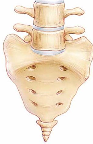
The sacral opening has individual dimensions
In the sacral spine, the elements are also connected motionless. Here, four or five vertebrae fused into a monolithic bone of a triangular shape with the apex pointing down. The sacrum is the base of the entire mobile spine, and has its own small amplitude of movement - up to 5 mm in the young years of man. It has two upper articular processes that are turned back and slightly to the sides. The sacrum is concave in the front, and the sacral and articular crest in the back, where there is an opening in the sacral canal, the sizes of which are very different for different people.
The structure of the lumbar vertebra differs from other similar elements in the massiveness of the “body”. From the first to the fourth element in the lower back, the vertebrae increase in size, and the fifth, last, takes part in the formation of an additional joint for connection with the upper part of the sacrum. The fifth, lower vertebra in the lower back, has not a classical cylindrical, but a wedge-shaped body. It is worth noting that in the lumbar articular processes at the top of the vertebrae are concave and face down and to the middle.
There are pits in the thoracic vertebrae
What is interesting about such an element of the skeleton as the thoracic vertebra? The structure here has such a feature - the presence on the "body" of the pits and half-holes for attaching the ribs. In addition, the vertebrae in the chest are larger than the cervical, but smaller than the lumbar, the height of the “bodies” increases gradually from the first to the twelfth.
It is also worth considering that the articular processes are located frontally, and the transverse processes are directed back and laterally. A remarkable feature of this section of the skeleton is that the spinous processes are inclined downward and overlap each other as in a tile. Each thoracic vertebra, the structure of which is shown in the figure, along with vertebrae from other departments is involved in such functions: creating support for the body, cushioning, protection. It contributes to the implementation of motor functions, takes part in metabolic and hematopoietic processes.
Among the cervical vertebrae there are Axis and Atlas
The structure of the cervical vertebrae is so different from the structure of these elements in other parts of the spine that two of them are even given individual names. The first is Atlas, the vertebra to which the human skull is attached. It does not have a “body”, instead of which there are two lateral “masses” connected by the front and rear arcs with the same tubercles. Atlant's lateral masses are provided with upper and lower articular surfaces, and on the posterior surface of the anterior arch there is a fossa to connect to the second vertebra - Axis. It is interesting that between the first vertebra and the skull there is no intervertebral disc, usually bearing a cushioning function.
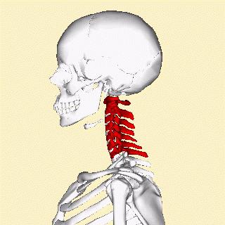
Axis in its structure has a "tooth" that enters the fossa in Atlanta, as well as the lower articular process and the spinous process (in contrast to Atlanta). The structure of the cervical vertebrae from the third to the sixth is a classic one with a pronounced "sleepy" tubercle on the transverse process of the sixth vertebra. The carotid artery is often pressed against this tubercle when bleeding must be stopped. The seventh vertebra in the cervical part has a long (non-bifurcated) process (spinous), which is why it is called a protruding vertebra, since it is guided by health workers when counting the vertebrae during the examination of the patient. The structural features of the cervical vertebrae are such that these elements have openings in the transverse processes, forming a bone channel through which large blood vessels pass to the brain, which provide nutrition to the most important organ in the human body.