The thoracic duct of the lymphatic system is its main vessel. It can be formed in several ways. Consider in detail what the thoracic duct is.
Anatomy
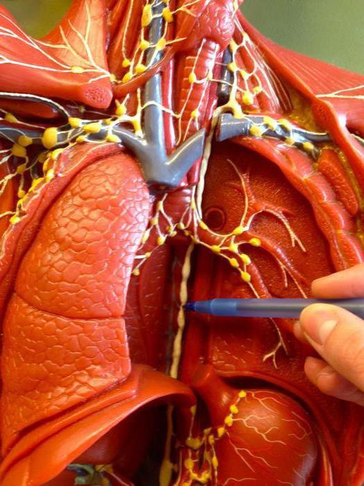
Three membranes are distinguished in the vessel wall: endothelial, muscular-fibrous, and external. In the first there are 7-9 large lunar valves. The muscular-fibrous membrane has a sphincter at the mouth. The adventitious (outer) part grows to the pleura, aorta, and spine. From the beginning in the duct secrete the abdominal, thoracic, cervical. The latter is presented in the form of an arc, and the first two - in the form of a long clearly shaped vessel that accompanies the descending aorta. The abdominal part passes through the aortic fissure in the diaphragm into the chest cavity. Here, the thoracic duct runs along the left lateral plane of the lower vertebrae posterior to the descending aorta. Then he deviates closer to the esophagus. In the region of the 2-3rd thoracic vertebra, the duct comes out from under the esophagus (its left edge). Then, behind the common and subclavian arteries, it rises to the upper aperture. Next, the vessel goes around the left and right pleura. Here, forming an arch, the thoracic duct flows into the venous angle or the branches forming it - the brachiocephalic, subclavian, internal jugular. On this site in the vessel a lunar valve and sphincter are formed. The thoracic duct reaches 1-1.5 cm in length, in rare cases 3-4 cm.
Formation
The thoracic duct is formed:
- The fusion of intestinal, lumbar or both of those and other trunks of both sides.
- The formation of the branches of the milky cistern. In this case, the thoracic duct looks like an ampullar, conical expansion.
- Merging only intestinal and lumbar trunks.
The thoracic duct can also form as a reticular in the form of a large looped plexus of celiac, lumbar, mesenteric branches and efferent vessels.
The specifics of the structure
Often in the topography and structure manifests variability. In particular, it is noted:
- Doubling of the thoracic region or the formation of additional (one or more) ducts.
- Different options for interaction with pleura, aorta, deep cervical veins, esophagus.
- Falling into the venous (jugular) angle, brachiocephalic, subclavian veins with several or one trunk.
- Formation of an ampoule in front of the site of entry into the vessels.
- Inflows can flow into the jugular angle or the veins that make it up independently.
Thoracic duct: right lymphatic duct
This element can also be formed in various ways:
- The merger of the subclavian, jugular, broncho-mediastinal trunks. In this case, a short and wide thoracic duct is formed . This situation is noted in 18-20% of cases.
- The right duct may be absent altogether. The trunks that form it directly open into the jugular angle or the vessels that make it up. This situation is observed in 80-82% of cases.
- There is a separation of a very short, wide right duct before entering the corner into 2-3 or more stems. This form of opening is called reticular.
Trunks
There are three of them:
- Jugular trunk. It is formed by efferent cervical vessels. They emerge from the deep and lateral nodes. This trunk accompanies the jugular internal vein to an angle. In this area, he flows into it or the vessels that form it, or takes part in the formation of the right duct.
- Subclavian trunk. Its occurrence is due to the fusion of efferent vessels from the axillary nodes. The trunk passes near the subclavian vein, has a sphincter and valves. It opens either into the venous corner and the vessels that form it, or into the right duct.
- Bronchomediastinal trunk. It is formed by efferent vessels from bronchopulmonary, tracheal, mediastinal nodes. There are valves in this barrel. It opens into the right duct, or venous jugular angle, or into the vessels that form it. The latter include the brachiocephalic, subclavian, jugular veins.
Left efferent vessels open in the thoracic duct. From the upper tracheobronchial and mediastinal nodes, they can flow into the venous corner. In the lymphatic trunks, as well as in the duct, three membranes are distinguished: adventitial, muscular-elastic and endothelial.
Pulmonary vessels and nodes
Capillaries form two networks. One - superficial - is located in the visceral pleura. The second - deep - is formed near the pulmonary lobules and alveoli, around the branches of blood vessels and the bronchial tree. The surface network is represented by a combination of narrow and wide capillaries. It is single layer. The capillaries are presented in the form of a plexus and spread over all surfaces in the visceral pleura. The deep web is three-dimensional. The lobular plexus is the main part of it. They send lymph in 2 directions. It enters the plexus of the pulmonary vessels and bronchi, as well as the pleural network. The bringing branches are formed at the level of segments, pass into the portal and lobar. They exit the lungs with veins and open into the following visceral nodes:
- Bronchopulmonary. They are divided into intraorgan and extraorgan. The former are located in the lobar and segmental bronchi, the latter in the root of the lung.
- Tracheobronchial upper and lower. They lie above and below the tracheal bifurcation.
The efferent vessels flow into the anterior mediastinal and tracheobronchial nodes. Of these, they open into the bronchomediastinal trunk. In rare cases, vessels can flow into the thoracic duct and jugular venous angle.