The hearing organ performs a function that is of great importance for the full functioning of a person. Therefore, it makes sense to study its structure in more detail.
Ear anatomy
The anatomical structure of the ears, as well as their components, has a significant impact on the quality of hearing. A person’s speech directly depends on the full operation of this function. Therefore, the healthier the ear, the easier it is for a person to carry out the process of life. It is these features that determine the fact that the correct anatomy of the ear is of great importance.
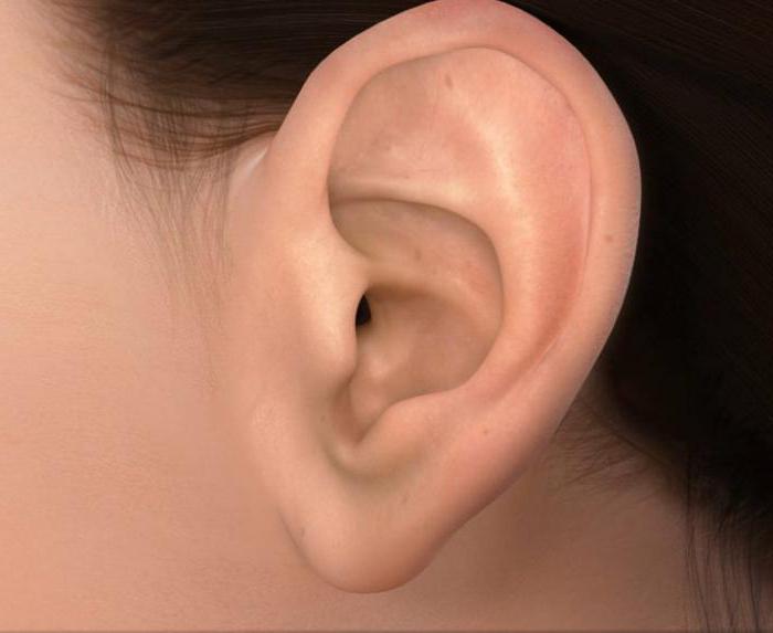
Initially, it is worthwhile to consider the structure of the organ of hearing from the auricle, which is the first that catches the eye of those who are not sophisticated in the subject of human anatomy. It is located between the mastoid process on the back side and the temporal mandibular joint in front. Thanks to the auricle, the perception of sounds by humans is optimal. In addition, it is this part of the ear that has important cosmetic value.
As the basis of the auricle, you can determine the cartilage plate, the thickness of which does not exceed 1 mm. On both sides it is covered with skin and perichondrium. The anatomy of the ear also indicates the fact that the only part of the shell devoid of the cartilaginous skeleton is the lobe. It consists of skin-covered fatty tissue. The auricle has a convex inner part and a concave outer one, the skin of which is tightly fused with the perichondrium. Speaking about the inner part of the shell, it is worth noting that in this area the connective tissue is much more developed.
The auricle is attached to the zygomatic, mastoid process and scales of the temporal bone through muscles and ligaments.
Anatomy of the outer ear
The external auditory meatus can be defined as the natural extension of the shell cavity. Its length in an adult is approximately 2.5 cm. In this case, the diameter can vary from 0.7 to 0.9 cm. This part of the ear has the shape of an epileptic or round lumen. The external part of the auditory meatus can be divided into two main departments: the external membranous-cartilaginous and the internal bone. The latter goes right up to the eardrum, which, in turn, delimits the middle and outer ears.
It is worth noting the fact that two-thirds of the length of the external auditory meatus is occupied by the membranous-cartilaginous section. As for the bone department, he gets only the third part. The continuation of the cartilage of the auricle, which has the form of an open back trough, acts as the basis of the membrano-cartilaginous department. Its cartilaginous skeleton is interrupted by upright santorinium cracks. They are closed with fibrous tissue. The border of the ear canal and the parotid salivary gland is located exactly at the place where these gaps are located. This fact explains the possibility of developing a disease that appeared in the outer ear, in the parotid gland. It should be understood that this disease can spread in the reverse order.
Those for whom the information on the topic “ear anatomy” is relevant should also pay attention to the fact that the membrano-cartilaginous section is connected to the bony part of the external auditory canal through fibrous tissue. The narrowest part can be found in the middle of this department. It is called the isthmus.
Within the membrano-cartilaginous region, the skin contains sulfur and sebaceous glands, as well as hair. It is from the secretion of these glands, as well as the scales of the epidermis that have been torn off, that earwax is formed.
Walls of the external auditory meatus
The anatomy of the ears includes information about the various walls that are located in the outer passage:
- The upper bone wall. If a fracture occurs in this part of the skull, then its consequence may be cerebrospinal fluid and bleeding from the ear canal.
- Front wall. It is located on the border with the temporomandibular joint. The transmission of movements of the jaw itself goes to the membranous-cartilaginous part of the external passage. Sharp painful sensations can accompany the chewing process if inflammatory processes are present in the area of the anterior wall.
- The anatomy of the human ear concerns the study and the posterior wall of the external auditory meatus, which separates the latter from the mast cells. At the base of this particular wall is the facial nerve.
- Bottom wall. This part of the external passage separates it from the salivary parotid gland. Compared to the top, it is 4-5 mm longer.
Hearing innervation and blood supply
These functions must be paid attention without fail to those who study the structure of the human ear. The anatomy of the organ of hearing includes detailed information about its innervation, which is carried out through the trigeminal nerve, the auricle of the vagus nerve, as well as the cervical plexus. Moreover, it is the posterior ear nerve that provides the nerves of the rudimentary muscles of the auricle, although their functional role can be defined as quite low.
Touching upon the topic of blood supply, it is worth noting that blood supply is provided from the external carotid artery system.
Blood supply directly to the auricle is carried out using the superficial temporal and posterior ear arteries. It is this group of vessels together with the branch of the maxillary and posterior ear arteries that provide blood flow in the deep parts of the ear and the eardrum in particular.
Cartilage is powered by vessels located in the perichondrium.
Within the framework of a topic such as “Anatomy and physiology of the ear”, it is worth considering the process of venous outflow in this part of the body and the movement of lymph. Venous blood leaves the ear along the posterior ear and posterior-mandibular veins.
As for the lymph, its outflow from the outer ear is carried out through nodes that are located in the mastoid process in front of the tragus, as well as under the lower wall of the auditory external passage.
Eardrum
This part of the organ of hearing performs the function of separating the outer and middle ear. In fact, we are talking about a translucent fibrous plate, which is strong enough and resembles an oval shape.
Without this plate, the ear cannot function fully. Anatomy reveals the structure of the tympanic membrane in sufficient detail: its size is approximately 10 mm, and its width is 8–9 mm. An interesting fact is that in children this part of the organ of hearing is almost the same as in adults. The only difference comes down to its shape - at an early age it is rounded and significantly thicker. If we take the axis of the external auditory canal as a reference, then the eardrum is obliquely relative to it, at an acute angle (approximately 30 °).
It is worth noting that this plate is located in the groove of the fibrous-cartilaginous drum ring. Under the influence of sound waves, the eardrum begins to tremble and transfers vibrations to the middle ear.
Drum cavity
The clinical anatomy of the middle ear includes information about its structure and functions. This part of the organ of hearing includes the tympanic cavity, as well as the auditory tube with a system of air cells. The cavity itself is a slit-like space in which 6 walls can be distinguished.
Moreover, in the middle ear there are three ear bones - an anvil, a malleus and a stapes. They connect with the help of small joints. In this case, the malleus is in close proximity to the eardrum. It is he who is responsible for the perception of sound waves transmitted by the membrane, under the influence of which the hammer begins to tremble. Subsequently, the vibration is transmitted to the anvil and stapes, and then the inner ear reacts to it. Such is the anatomy of the human ears in their middle part.
How the inner ear works
This part of the hearing organ is located in the temporal bone and looks like a labyrinth. In this part, the resulting sound vibrations are converted into electrical impulses that are sent to the brain. Only after the completion of this process is a person able to respond to sound.
It is important to pay attention to the fact that the inner ear of a person contains semicircular canals. This is relevant information for those who study the structure of the human ear. The anatomy of this part of the organ of hearing has the form of three tubes that are curved in the shape of an arc. They are located in three planes. Due to the pathology of this ear department, disturbances in the functioning of the vestibular apparatus are possible.
Sound anatomy
When the energy of sound enters the inner ear, it is converted into impulses. Moreover, due to structural features of the ear, a sound wave propagates very quickly. The consequence of this process is the occurrence of hydrostatic pressure, contributing to the shift of the integumentary plate. As a result, deformation of stereocilia of hair cells occurs, which, having come to a state of excitement, transmit information using sensory neurons.
Conclusion
It is easy to see that the structure of the human ear is quite complex. For this reason, it is important to ensure that the organ of hearing remains healthy and to prevent the development of diseases found in this area. Otherwise, you may encounter a problem such as impaired sound perception. To do this, at the first symptoms, even if they are insignificant, it is recommended to pay a visit to a highly qualified doctor.