The heart is the main organ of the human body. It is a muscular organ hollow inside and shaped like a cone. In newborns, the heart weighs about thirty grams, and in an adult - about three hundred.
The topography of the heart is as follows: it is located in the chest cavity, moreover, its third part is located on the right side of the mediastinum, and two thirds - on the left. The base of the organ is directed upward and somewhat posteriorly, and the narrow part, that is, the apex, is directed downward, to the left and anteriorly.
Organ boundaries
The boundaries of the heart allow you to determine the location of the organ. There are several of them:
- Top. It corresponds to the cartilage of the third rib.
- Lower. This border connects the right side with the apex.
- Top. This border is located in the fifth intercostal space, in the direction of the left midclavicular straight line.
- The right one. Between the third and fifth rib, a couple of centimeters to the right of the edge of the sternum.
- Left. The topography of the heart at this border has its own characteristics. It connects the apex with the upper border, and it passes along the left ventricle, which is facing the left lung.
According to topography, the heart is behind and just below half of the sternum. The largest vessels are located behind, in the upper part.
Topography Changes
The topography and structure of the heart in a person changes with age. In childhood, the body makes two turns around its axis. The borders of the heart change during breathing and depending on the position of the body. So, in lying on the left side and when bending, the heart approaches the chest wall. When a person is standing, it is lower than when he is lying. Due to this feature, the apical impulse is displaced. By anatomy, the topography of the heart also changes as a result of respiratory movements. So, on inspiration, the organ moves farther from the chest, and on exhalation it comes back.
Changes in the function, structure, and topography of the heart are observed in different phases of cardiac activity. These indicators depend on gender, age, as well as on the individual characteristics of the body: the location of the digestive system.
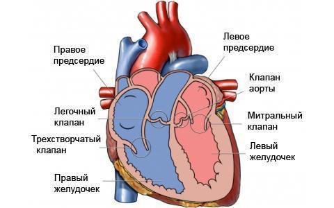
Heart structure
The heart has a top and bottom. The latter is turned up, to the right and back. At the back, the base is formed by the atria, and in the front, by the pulmonary trunk and the large artery, the aorta.
The top of the organ is facing down, forward and to the left. According to the topography of the heart, it reaches the fifth intercostal space. The apex is usually located at a distance of eight centimeters from the mediastinum.
The walls of the organ have several layers:
- Endocardium.
- Myocardium.
- Epicardium.
- Pericardium.
The endocardium is lined with an organ inside. This fabric forms valves.
The myocardium is a cardiac muscle that involuntarily contracts. The ventricles and atria also consist of muscles, and in the former the muscles are more developed. The surface layer of the muscles of the atria consists of longitudinal and circular fibers. They are independent for each atrium. And in the ventricles there are the following layers of muscle tissue: deep, superficial and middle circular. From the deepest fleshy lintels and papillary muscles are formed.
The epicardium is epithelial cells covering the outer surface and organ and the nearest vessels: the aorta, vein, and also the pulmonary trunk.
The pericardium is the outer leaf of the pericardial sac. Between the leaves there is a slit-like formation - the pericardial cavity.
Holes
The heart has several holes, chambers. The organ has a longitudinal septum, which divides it into two parts: left and right. Atria are located at the top of each part, and ventricles are at the bottom. There are holes between the atria and ventricles.
The first of them have some protrusion, which forms the heart ear. The walls of the atria have different thicknesses: the left is more developed than the right.
There are papillary muscles inside the ventricles. Moreover, there are three in the left, and two in the right.
In the right atrium, the fluid comes from the superior and inferior genital veins, sinus veins. Four pulmonary veins lead to the left . The pulmonary trunk leaves from the right ventricle , and the aorta from the left ventricle.
Valves
In the heart there are tricuspid and bicuspid valves, which close the gastro-atrial openings. The absence of reverse blood flow and eversion of the walls is provided by tendon filaments passing from the edge of the cusps to the papillary muscles.
A bicuspid or mitral valve closes the left ventricular-atrial orifice. Tricuspid - right ventricular-atrial foramen.
In addition, there are lunar valves in the heart . One closes the aortic opening, and the other closes the pulmonary trunk. Valve defects are defined as heart disease.
Circulatory Circles
In the human body there are several circles of blood circulation. Consider them:
- The large circle (BCC) starts from the left ventricle and ends with the right atrium. Blood flows through it through the aorta, then through the arteries, which diverge into precapillaries. After this, the blood enters the capillaries, and from there to the tissues and organs. In these small vessels, nutrients are exchanged between tissue cells and blood. After this, the reverse flow of blood begins. From capillaries, it enters post-capillaries. They form venules, from which venous blood enters the veins. According to them, she approaches the heart, where the vascular channels converge into the vena cava and enter the right atrium. So there is a blood supply to all organs and tissues.
- Small circle (MCC) starts from the right ventricle and ends with the left atrium. Its beginning is the pulmonary trunk, which is divided into a pair of pulmonary arteries. Venous blood flows through them. It enters the lungs and is enriched with oxygen, turning into arterial. Blood then collects into the pulmonary veins and flows into the left atrium. MCC is intended to enrich the blood with oxygen.
- There is still a crown ring. It starts from the aortic bulb and the right coronary artery, passes through the capillary network of the heart and returns through the venules and coronary veins, first to the coronary sinus, and then to the right atrium. This circle supplies the heart with nutrients.
The heart, as you can see, is a complex organ with its own circulation. Its boundaries change, and the heart itself changes its angle of inclination with age, turning around its axis twice.