The stomach belongs to the main organs of the digestive tract. It processes all the products that we consume. This is due to hydrochloric acid, which is present in the stomach. This chemical compound is secreted by special cells. The structure of the stomach is represented by several types of tissues. In addition, cells that secrete hydrochloric acid and other biologically active substances are not located throughout the organ. Therefore, the anatomically stomach consists of several sections. Each of them differs in functional value.
Stomach: organ histology
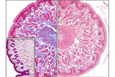
The stomach is a hollow organ of a bag-shaped form. In addition to chemical treatment of chyme, it is necessary for the accumulation of food. To understand how digestion is carried out, you should know what the histology of the stomach is. This science studies the structure of organs at the tissue level. As you know, living matter consists of many cells. They, in turn, form tissues. The cells of the body are different in structure. Therefore, the fabrics are also different. Each of them performs a specific function. The internal organs are composed of several types of tissue. Thanks to this, their activities are ensured.
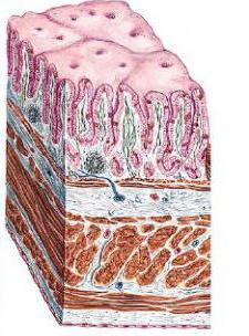
The stomach is no exception. Histology studies 4 layers of this organ. The first of these is the mucous membrane. It is located on the inner surface of the stomach. Further there is a submucosal layer. It is represented by adipose tissue, in which there are blood and lymph vessels, as well as nerves. The next layer is the muscularis. Thanks to it, the stomach can contract and relax. The last is the serous membrane. She is in contact with the abdominal cavity. Each of these layers consists of cells that together form tissue.
Histology of the gastric mucosa
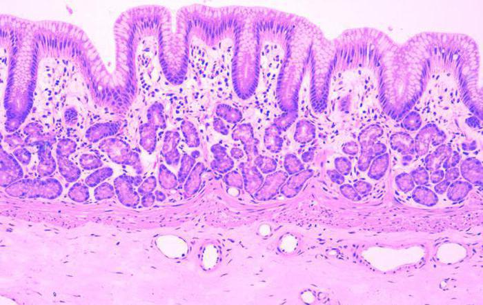
The normal histology of the gastric mucosa is represented by epithelial, glandular and lymphoid tissue. In addition, in the composition of this shell there is a muscle plate, consisting of smooth muscles. A feature of the gastric mucosa is that there are many pits on its surface. They are located between the glands secreting various biological substances. Next is a layer of epithelial tissue. It is followed by the gland of the stomach. Together with lymphoid tissue, they form their own plate, which is part of the mucous membrane.
Glandular tissue has a certain structure. It is represented by several entities. Among them:
- Simple glands. They have a tubular structure.
- Branched glands.
The secretory section consists of several exo- and endocrinocytes. The excretory duct of the glands of the mucous membrane enters the bottom of the fossa, located on the surface of the tissue. In addition, cells in this section are also able to secrete mucus. The gaps between the glands are filled with coarse connective fibrous tissue.
Lymphoid elements may be present in the lamina propria mucosa. They are located diffusely, but the entire surface. This is followed by muscle plate. It consists of 2 layers of circular fibers and 1 - longitudinal. He occupies an intermediate position.
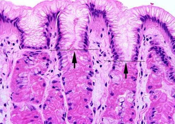
The histological structure of the epithelium of the stomach
The upper layer of the mucous membrane, which is in contact with food masses, is the epithelium of the stomach. The histology of this gastrointestinal tract is different from the structure of the tissue in the intestine. The epithelium not only protects the surface of the organ from damage, but also has a secretory function. This tissue lines the cavity of the stomach from the inside. It is located on the entire surface of the mucous membrane. No exception and gastric fossa.
The inner surface of the organ is covered with a single-layer prismatic glandular epithelium. The cells of this tissue are secretory. They are called exocrinocytes. Together with the cells of the excretory ducts of the glands, they produce a secret.
Histology of the fundus of the stomach
The histology of different parts of the stomach is not the same. Anatomically, the organ is divided into several parts. Among them:
- Cardiac department. At this point, the esophagus passes into the stomach.
- Bottom. In another way, this part is called the fundal department.
- The body is represented by large and small curvature of the stomach.
- Antral department. This part is located before the transition of the stomach into the duodenum.
- Pyloric department (pylorus). In this part, there is a sphincter connecting the stomach to the duodenum. The gatekeeper occupies an intermediate position between these bodies.
Of great physiological importance is the fundus of the stomach. The histology of this area is complex. The fundus has its own glands of the stomach. Their number is about 35 million. The depth of the fossa between the fundus glands occupies 25% of the mucous membrane. The main function of this department is the production of hydrochloric acid. Under the influence of this substance, biologically active substances (pepsin) are activated, food is digested, and the body is protected from bacterial and viral particles. Own (fundamental) glands consist of 2 types of cells - exo and endocrinocytes.
Histology of the submucous membranes of the stomach
As in all organs, under the mucous membrane of the stomach is a layer of adipose tissue. Vascular (venous and arterial) plexuses are located in its thickness. They supply blood to the inner layers of the wall of the stomach. In particular, the muscle and submucous membranes. In addition, in this layer there is a network of lymphatic vessels and a nerve plexus. The muscular membrane of the stomach is represented by three layers of muscle. This is a distinctive feature of this body. Outside and inside are longitudinal muscle fibers. They have an oblique orientation. Between them lies a layer of circular muscle fibers. As in the submucosa, there is a nerve plexus and a network of lymphatic vessels. Outside, the stomach is covered with a serous layer. It is a visceral peritoneum.
Benign neoplasms of the stomach and intestines: histology of hemangiomas
One of the benign neoplasms is hemangioma. The histology of the stomach and intestines with this disease is necessary. Indeed, despite the fact that the education is benign, it should be differentiated from cancer. Histologically, hemangioma is represented by vascular tissue. The cells of this tumor are completely differentiated. They do not differ from the elements that make up the arteries and veins of the body. Most often, hemangioma of the stomach is formed in the submucosal layer. The pyloric department is considered a typical localization for this benign neoplasm. The tumor may have various sizes.
In addition to the stomach, hemangiomas can be localized in the small and large intestines. These formations rarely make themselves felt. However, the diagnosis of hemangiomas is important. With large sizes and constant trauma (chyme, feces), serious complications can occur. The main one is profuse gastrointestinal bleeding. A benign neoplasm is difficult to suspect, since in most cases there are no clinical manifestations. An endoscopic examination reveals a dark red or cyanotic rounded spot that rises above the mucous membrane. In this case, the diagnosis of hemangioma is made. The histology of the stomach and intestines is crucial. In rare cases, hemangioma undergoes malignant degeneration.
Stomach Regeneration: Histology for Ulcer Healing
One of the indications for histological examination is gastric ulcer. With this pathology, an endoscopic examination (FEGDS) is performed with a biopsy taken. Histology is mandatory for suspected malignancy of an ulcer. Depending on the stage of the disease, the resulting tissue may be different. When healing an ulcer, a scar of the stomach is examined. Histology in this case is needed only if there are symptoms due to which it is possible to suspect malignant degeneration of the tissue. If there is no malignancy, then cells of coarse connective tissue are detected in the analysis. With malignancy of a stomach ulcer, the histological picture may be different. It is characterized by a change in the cellular composition of the tissue, the presence of undifferentiated elements.
What is the purpose of histology of the stomach?
One of the organs of the digestive tract, in which tumors often develop, is the stomach. Histology should be performed in the presence of any change in the mucous membrane. The following diseases are considered indications for this study:
- Atrophic gastritis. This pathology is characterized by depletion of the cellular composition of the mucous membrane, inflammation, and a decrease in the secretion of hydrochloric acid.
- Rare forms of gastritis. These include lymphocytic, eosinophilic and granulomatous inflammation.
- Chronic peptic ulcer of the stomach and duodenum.
- The development of "small signs" according to Savitsky. These include general weakness, decreased appetite and performance, weight loss, a feeling of discomfort in the abdomen.
- Detection of polyps of the stomach and other benign neoplasms.
- A sudden change in the clinical picture with long-term peptic ulcer. These include a decrease in the intensity of the pain syndrome, the development of aversion to meat food.
The listed pathologies relate to precancerous diseases. This does not mean that the patient has a malignant tumor, and its location is the stomach. Histology helps determine exactly what changes are observed in the tissues of the organ. To prevent the development of malignant degeneration, it is worthwhile to conduct a study as early as possible and take measures.
The results of histology of the stomach
Histological findings may vary. If the organ tissue is not changed, then microscopy reveals a normal prismatic single-layer glandular epithelium. When taking a deeper layer for a biopsy, you can see smooth muscle fibers, adipocytes. If the patient has a scar from a prolonged ulcer, a rough fibrous connective tissue is detected. With benign formations, the results of histology may be different. They depend on what tissue the tumor evolved from (vascular, muscle, lymphoid). The main feature of benign formations is cell maturity.
Stomach tissue sampling for histology: methodology
To perform a histological examination of the tissue of the stomach, it is necessary to perform an organ biopsy. In most cases, it is performed using endoscopy. The apparatus for performing PEGF is placed in the lumen of the stomach and several pieces of organ tissue are cleaved. It is advisable to take biopsy samples from several remote sites. In some cases, tissue for histological examination is taken during surgery. After that, thin sections from the biopsy specimen are examined in the laboratory and examined under a microscope.
How long does a histological analysis of stomach tissue take
If cancer is suspected, histology of the stomach is necessary. How long does this analysis do? Only the attending physician can answer this question. On average, histology takes about 2 weeks. This applies to planned studies, for example, when removing a polyp.
During the operation, an urgent histological examination of the tissue may be necessary. In this case, the analysis takes no more than half an hour.
In which clinics is a histological analysis performed?
Some patients are interested in: where can I urgently make histology of the stomach? This study is carried out in all clinics that have the necessary equipment and laboratory. Urgent histology is carried out in oncology clinics, some surgical hospitals.