In this article, we will examine the function of the rectum and its significance. And also get acquainted with its anatomical structure, analyze in detail the role of the layers of which it consists, study the processes of blood supply.
General information about the rectum
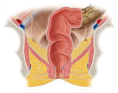
The rectum is necessary for the body to accumulate feces. It originates in the area of the cape, then descends into the cavity of the small pelvis, located in front of the sacrum. This structure forms 2 bends, moving from front to back and referred to as upper and lower. The upper one is convex in the direction of concavity of the sacrum, and the lower one looks into the coccyx. Sometimes it is called perineal.
The upper section and the end
Features of the structure and functions of the rectum depend primarily on its constituent elements, departments, cells and location. One of these components is the upper part of the organ and its final part.
The upper section is a kind of ampoule, the diameter of which is usually in the range of 8-16 cm, but this number can increase due to, for example, atony. This formation is located in the pelvic cavity and expands at one end.
The final part is represented by a circle directed down and back, and its continuation is located in the anal canal. After passing the pelvic floor ends with a hole. The sizes of the formed circle vary less than that of the upper section, and correspond to 5–9 cm. The size of the intestine ranges from 13 to 16 cm, but about 65–85% of it falls into the pelvic section, and the remaining centimeters form the anal section.
The structure of the mucous membrane
The functions of the human rectum are largely determined by its mucous membrane. The mucous membrane forms a large number of longitudinal folds, which is possible due to its developed submucosa. These folds can be easily smoothed out due to stretching of the intestinal wall. The anal canal has folds with a constant look; there are eight to ten of them. These formations have special depressions lying between them, and are called anal sinuses (clinicians), which are clearly expressed in children. It is the clinicians who accumulate in themselves a special mucus that facilitates the passage of feces through the anal canal. Anal sinuses are also called anal crypts. They most often serve as the front door for microorganisms. The tissue layer, located between the anus and sinuses, includes a plexus of veins. In addition to the longitudinal folds, the upper sections of the rectum have transverse folds. These formations are very similar to the lunate folds of the sigmoid colon.
Muscle Shell Description
The structure and functions of the rectum also depend and are determined by the muscular membrane, which consists of 2 layers, namely: circular and longitudinal. The circular (inner) layer begins to thicken in the upper section of the perineal region. It is in this area that the internal sphincter forms, which ends in the area of the junction of the skin and anal canal. The longitudinal layer covers both the anterior and posterior parts of the intestine, equivalently. In the lower part, the longitudinal fiber begins to intertwine with the muscle that rises towards the anus, and is also often intertwined with the external sphincter.
Thanks to this, we can conclude that the rectum has the features of the conducting section of the digestive canal and is similar to the esophagus. There is a similarity between these structures in the development process: both ends of the primary intestine during embryogenesis undergo a breakthrough in the blind end of the tube. In the esophagus, this occurs with the pharyngeal membrane, and in the rectum with the cloacal membrane. Both channels have musculature consisting of two continuous layers.
Topographic information
The functions of the rectum can be described with topographic information. Behind the organ are two divisions of the spine, sacral and coccygeal. And in front of the male, the intestine is adjacent to the seminal vesicles and the vas deferens. The rectum of women borders in the anterior section with the posterior vaginal wall and uterus. It is separated from these structures by a layer formed by connective tissue.
Own fascia of the rectum and the anterior surface of the sacral and coccygeal spine do not have fascial jumpers. This simplifies the operation to remove the intestine and its fascia, covering the vessels. Doctors have no particular problems with this.
The functions of the rectum. Description
One of the functions of the rectum is to retain food residues that have not had time to absorb in the area of the cavity of the small intestine, as well as water. This includes a large number of substances of an organic nature and products that have undergone bacterial decay, and also contains substances that cannot be digested, such as fiber. There is also bile, bacterial organisms, salts.
In connection with the functions of the rectum, processes such as the breakdown of food that is not digested in other parts of the food department are observed there. And the formation of feces. In the colon, the secretion of digestive juice constantly occurs, containing the same set of enzymes as in the small intestine, but with a less pronounced effect. Here, the collection of gases takes place.
The key function of the rectum is the removal of waste from the life process. Or, in other words, the excretion of feces from the body. Mostly this process is regulated by the consciousness and will of man.
Violation of the function of the rectum, as a rule, is the result of a sedentary way of life, poor nutrition, neuro-emotional overload, etc. Most often, such stressful situations lead to constipation. To a violation of the intestines, which affects the bowel movement.
Blood circulation processes
The blood supply to the rectum is due to the unpaired upper rectal and two paired rectal. A well-developed network of vessels of the sigmoid colon makes it possible to maintain an unpaired rectal artery, namely, its marginal vessels, full blood supply even due to high intersections of the rectal paired arteries and the sigmoid.
The middle paired arteries emerging from the branch of the iliac arteries sometimes develop in different ways, and, sometimes, are absent. And, nevertheless, in some situations they can play a key role in the blood supply process.
The lower arteries, originating from the internal sacral artery, supply the external sphincter and skin. Plexuses of veins are located in a wide variety of layers of the intestinal wall. Among them are:
- submucosal plexus - has a circular shape, consists of a submucous membrane and venous trunks, and is also associated with the other two plexuses;
- subfascial plexus;
- subcutaneous plexus.
Finally
In short, the function of the human rectum can be summarized as follows. This body is responsible, first of all, for the place of storage of feces and the reservoir for the accumulation of gas. Also here is the breakdown of undigested food and the removal of waste from the life process.