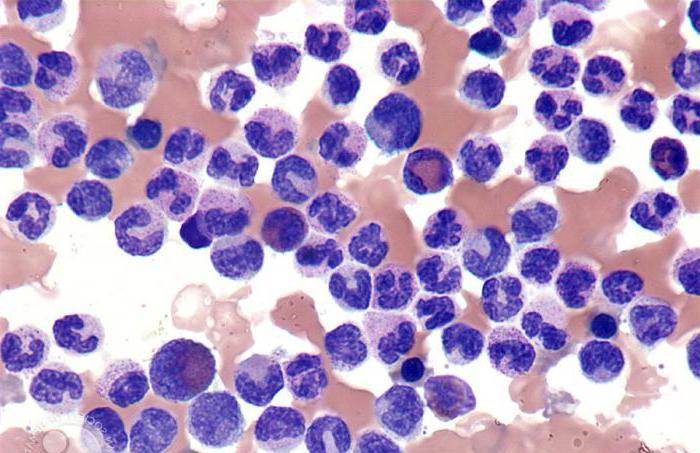
Leukemoid reactions - changes in hematopoiesis, similar to the blood picture in leukemia and other tumors of the hematopoietic system. It should be noted that the specificity of these effects is considered to be their active orientation and the lack of transition to oncological pathology. These reactions can be triggered by various types of intoxications, tumors, infections, metastases of brain cancer.
The development mechanism is not the same for different types of reactions: in some cases, it is the release of immature cellular elements into the blood, in others it is increased production of blood cells or restriction of the release of cells into tissues, or the presence of several mechanisms simultaneously.
What could be the source of the disease?
There are many factors that may cause leukemoid reactions. The reasons for their development are as follows:
- the effect of ionizing radiation;
- tuberculosis;
- sepsis;
- purulent processes;
- dysentery;
- lymphogranulomatosis;
- skull injuries;
- shock state;
- lobar pneumonia;
- erysipelas;
- diphtheria;
- scarlet fever;
- acute liver dystrophy;
- corticoid hormone therapy;
- carbon monoxide poisoning.
Types of disease
The following types of leukemoid reactions are distinguished:
- Myeloid reactions.
- Lymphocytic.
- Pseudoblasts.
Let's consider each of them in more detail.
Myeloid
This type includes reactions such as neutrophilic, promyelocytic and eosinophilic. Leukemoid effects, similar to chronic myelogenous leukemia, are accompanied by intoxication and severe infections. Active leukocytosis at its core always has a complex process, accompanied by the presence of sepsis, inflammatory foci and an increase in body temperature.
Exposure to excess eosinophils in the blood, as a rule, occurs with sensitization to parasites and drugs, allergic diathesis, rarely with cancer (lymphogranulomatosis and lymphosarcoma). Leukemoid Reactions Data need a comprehensive examination to eliminate diseases of the circulatory system and helminths.
Reactive cells are similar to erythremia. Erythrocytosis factors are often lung ailments with a decrease in blood oxygenation (oxygen saturation), kidney tumors, as well as congenital heart defects. In this situation, computer and ultrasound are required.
Myelia is similar to acute erythromyelosis, which differs only in the lack of blast red blood cells in the bone marrow and blood. Often it can be detected with bone metastases of the disease.
Lymphocytic
Such reactions are characterized by a significant increase in the total number of lymphocytes in the peripheral blood, which is often responsible for an increase in the liver, lymph nodes and spleen.
This type includes mononucleosis, infectious lymphocytosis, monocyte-macrophage leukemoid reactions in children with bacterial, viral infections, as well as parasitic infections and large blood eosinophilia (for example, with helminthiases).
Lymphocytic reactions appear:
- with viral infections (chickenpox, rubella, mumps, adenovirus infection, measles, infectious mononucleosis);
- parasitic infections (rickettsiosis, toxoplasmosis, chlamydia);
- bacterial infections (syphilis, whooping cough, tuberculosis);
- different mycoses;
- autoimmune diseases (serum disease, systemic lupus erythematosus).
The lymphocytic type still occurs with Waldenstrom macroglobulinemia, inflammatory processes, sarcoidosis. All of the above ailments are treated very hard and can disturb the patient for more than one year.
Pseudoblast
Such leukemoid reactions occur if the patient is just starting to get out of immune agranulocytosis, the cause of which can be caused by taking sulfonamides, Amidopyrine, Butadion and other drugs.
Such a group of effects is characterized by the presence in the peripheral blood and bone marrow of a considerable number of cellular components with a homogeneous nucleus, single nucleoli and a blue, thin, non-granular cytoplasm.
In contrast to the characteristic blast erythrocytes, these cells do not have a specific soft network and dimension of chromatin fibers. Intermittent blastoses that disappear without chemotherapy and are related to leukemoid effects are found in newborn babies with genetic chromosome disorders (e.g., Down syndrome).
Leukemoid reactions, the types of which were presented above, forming against the background of any pathology, usually do not provoke dangerous complications. Sometimes a sharp thrombocytopenia can be mistakenly regarded as one of the signs of acute leukemia. In the detection of immunoblastic lymphadenitis, the safety of the natural structure of the lymph node, as well as the precisely defined lines of the follicles, are of considerable importance.
Leukemoid reactions and leukemia: differences
There are some differences between such effects and leukemia, namely:
- With leukemoid reactions, there is no rapid rejuvenation of the bone marrow, it is metamyelocytic, and with leukemia an increase in blast forms is detected. With leukemoid effects, the erythroid sprout is preserved, there is a normal leukoerythroblastic ratio of 3: 1 and 4: 1.
- Manifested anaplasia is not observed with leukemoid phenomena, as is the case with leukemia, when protoplasm bulges out and an anomaly of the nucleus occurs.
- In the first variant, the absolute number and an increase in the percentage of mature neutrophils are noted in peripheral blood, with leukemia, the content of mature neutrophils decreases, and excess proliferation of young, immature forms occurs.
- With leukemoid reactions, toxic granularity of neutrophils is often noted.
- During a cytochemical study of leukocytes with leukemia, a decrease or absence of alkaline phosphatase is observed , with leukemoid reactions an increased activity.
- With exacerbation of chronic myelogenous leukemia, the eosinophilic-basophilic association is a harbinger of blast crisis; in leukemoid reactions, it is absent.
- With myeloid leukemia, high thrombocytosis is often observed, with leukemoid reactions the platelet count is within normal limits.
- In the initial stages of chronic myelogenous leukemia, a large dense spleen is found, with leukemoid reactions, splenomegaly also sometimes occurs, but this organ is soft and never reaches very large sizes.
- With leukemoid reactions to a neoplastic process , cancer cells are found in the bone marrow.
Leukemoid reactions in children: diagnostic algorithm
An important role in the diagnosis of this disease is assigned to a pathologist who examines the biopsy material. But in order to prevent an irreparable mistake, the pathologist must collect reliable information about the patient, give him direction for various tests and prescribe a cytostatic treatment that will remove all the consequences of lymphadenitis. If all this is not done, then the diagnosis will be made incorrectly, and therefore it will be very difficult to cope with the disease. After all, such a disease is very dangerous. Sometimes, for biopsy details, a second biopsy is necessary.
Of considerable importance in the diagnosis is a smear from the exterior of the biopsy lymph node and an imprint. With lymphosarcoma, most of the red blood cells (at least 30 percent) are constant blast cells. In immunoblastic lymphadenitis, such red blood cells are usually less than 10 percent, they are different in terms of basophilia of the cytoplasm and the maturity of the nucleus.
The histopathological diagnosis of the analysis of the lymph node should be very detailed and exclude an inaccurate conclusion. Because the pathologist for various blood tests should clearly determine the diagnosis, and this is reflected in the conclusion. For example, in order to establish an initial diagnosis of benign lymphomas, in some cases it is necessary to observe the patient for a long time and examine the lymph nodes again.
Diagnosis of leukemoid reactions suspected of detecting monoclonal immunoglobulin sometimes requires years of follow-up and repeated bone marrow punctures. Until the diagnosis is approved, antitumor treatment is contraindicated.
Infectious mononucleosis
Also called Filatov's disease - Pfeiferra, glandular fever and monocytic tonsillitis. It is a viral disease characterized by blast transformation of lymphocytes, enlarged lymph nodes and spleen, reactive lymphadenitis, the appearance of specific red blood cells in peripheral blood. The causative agent is the Epstein-Barr virus. At the base of the disease is the blast transformation of lymphocytes caused by a special viral infection.
The clinical situation is different. In mild forms, health is disturbed due to rhinitis. Indicative signs:
- tonsillitis ("burning pharynx");
- an increase in the spleen and cervical lymph nodes, as well as their pain;
- difficult nasal breathing in the early days of the disease due to swelling of the mucous membrane.
Blood condition: increased percentage of eosinophils, lymphocytes and monocytes.
Complications
A necessary and sufficient indicator of the disease is the presence in the blood of a kind of mononuclear cells (over 10-20%) - cells that differ in the nucleus of a large lymphocyte and a broad basophilic cyanoplasm with a lilac color with pronounced perinuclear enlightenment. Leukemoid reactions in children and adults last for several weeks, but in certain situations, normalization of the blood condition lasts for months.
Relapses with a milder course are also observed, sometimes at intervals of several years after the first acute period. Complications may include:
- acute hepatitis;
- encephalitis;
- agranulocytosis;
- rupture of the spleen caused by its rapid increase;
- autoimmune hemolysis.
Therapy of leukemoid reactions
As a rule, patients do not need special medication, because within a few days the main signs of the disease disappear and the blood condition returns to normal. In case of a protracted illness and ill state of the patient, pathogenetic therapy is used - Prednisolone is prescribed in a dosage of 20-30 milligrams per day or other glucocorticoid drugs to eliminate leukemoid reactions. In any case, treatment is prescribed only by a specialist.
Forecast
Usually positive: contagiousness is small, and therefore patient quarantine is optional. However, spleen ruptures are very dangerous . Recovery is determined by the appearance of signs of a decrease in organ volumes, as well as the disappearance of tonsillitis and normalization of body temperature. If infectious mononucleosis manifests itself in the form of hepatitis, hospitalization of the patient is required.