Shin refers to the lower limb. It is located between the foot and the knee. Shin is formed by means of two bones - small and tibial. They are surrounded by muscle fibers on three sides. The tibial muscles, the anatomy of which will be discussed later, move the fingers and foot.
Tibia
This item has an extension on the top edge. Condyles are formed in this area: lateral and medial. On top of them are the surfaces of the joints. They articulate with the condyles of the thigh. On the lateral segment, there is an articular surface outside, through which there is a connection with the head in the fibula. The body of the tibial element looks like a trihedral prism. Its base is directed posteriorly and accordingly has 3 surfaces: rear, outer and inner. Between the last two is the edge. It is called the front. In its upper part, it passes into the tuberosity of the tibia. This area is designed to fix the tendon of the quadriceps femoris. In the lower part of the tibia has an extension, and on the inner surface there is a ledge. It is oriented downward. This protrusion is called the medial ankle. On the back side of the bone lies a rough piece of soleus muscle. The articular surface is located on the distal pineal gland. It serves to connect with the talus.
Second element
The fibula is thin, long, located laterally. Its upper end has a thickening - the head. It connects to the tibia. The lower part of the element is also thickened and forms a lateral ankle. She, like the head of the fibula, is oriented outwards and is well felt.
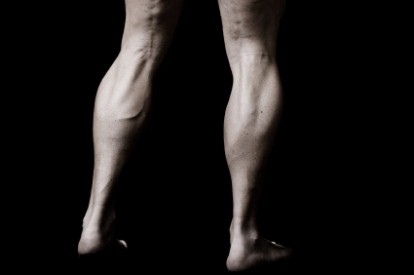
Shin muscles: their location, functions
Fibers are located on three sides. Different muscles of the leg are distinguished. The front group performs extension of the foot and fingers, supination and adduction of the foot. This segment includes three types of fibers. The tibial anterior tibialis muscle was the first to be formed. The remaining fibers form long extensors of the fingers and separate for the thumb on the foot. The back muscle group of the lower leg forms more fibers. In particular, there are long flexors of the fingers and separately - for the large, popliteal, triceps muscle of the leg. There are also tibial fibers. The short and long peroneal muscles of the lower leg are referred to the outer group. These fibers bend, penetrate and deflect the foot.
Tibial segment
This anterior muscle of the tibia begins from the eponymous bone, its external surface, fascia and interosseous membrane. They are directed down. The fibers pass under two bundles. They are located in the area of the ankle joint and ankles. These areas - the upper and lower extensor tendon retinaculi - are represented by thickening sites of the fascia of the foot and lower leg. The site of fiber attachment is the sphenoid medial and the base of the metatarsal (first) bone. The muscle is reasonably well felt throughout its entire length, especially at the site of transition to the foot. At this point, her tendon protrudes during extension. The task of this leg muscle is supination of the foot.
Finger extensor (long)
It runs from the anterior muscle outwards in the upper region of the lower leg. Its fibers begin from the head and marginal sections of the tibia, fascia and interosseous membrane. The extensor, moving to the foot, is divided into five tendons. Four are attached to the distal phalanges of the fingers (second to fifth), the latter to the base of the 5th metatarsal bone. The task of the extensor, acting as a multi-joint muscle of the leg, is not only to coordinate the extension of the fingers, but also the foot. Due to the fact that one tendon is fixed on its edge, the fibers also penetrate the area somewhat.
Thumb Extensors
Fibers begin in the region of the lower leg from the interosseous membrane and the inner part of the fibula. The extensors are less powerful than the segments described above. The attachment site of this is the distal phalanx in the thumbs. These leg muscles not only perform their extension, but also the feet, also contributing to their supination.
Finger flexor (long)
It starts from the back of the tibia, passing under the medial ankle to the foot. The channel for it is located under the retainer (ligament) of the flexor tendons. Next, the muscle is divided into four segments. On the foot (its plantar surface), the fibers cross the tendon from the flexor (long) thumb. Then they are joined by the square muscle of the sole. Four formed tendons are fixed to the distal phalanges (at their base) of 2-5 fingers. The task of this muscle is, among other things, in flexion and supination of the foot. Fiber square fibers are attached to the tendon. Due to this, averaging of muscle action occurs. Lying under the medial ankle and fan-shaped towards the phalanges, the long flexor also provokes some reduction of the fingers to the median surface of the body. Due to the pulling of the tendon by the square muscle, this action is slightly reduced.
Triceps muscle
It runs along the back surface and has 3 heads. Two form the surface area - the calf muscle, from the third - deep - fibers of the sole sole segment extend. All heads connect and form the common Achilles (calcaneal) tendon. It attaches to the tubercle of the corresponding bone. The calf muscle starts from the femoral condyles: lateral and medial. The task of two heads located in this area is twofold. They coordinate the flexion in the knee joint and the foot in the ankle. The medial element descends slightly lower and is better developed than the lateral one. The soleus muscle departs from the back side in the upper third of the tibia. It also attaches to the tendon arch located between the bones. The fibers extend slightly lower and deeper than the calf. They lie behind the subtalar and ankle joints and cause flexion of the foot. The triceps muscle can be felt under the skin. The calcaneal tendon protrudes posteriorly from the transverse axis in the ankle joint. Thanks to this, the triceps muscle has a large moment of rotation relative to this line. The heads of the calf segment participate in the formation of the rhomboid popliteal fossa. Its boundaries are: the biceps femoris (outside and above), the semi-membranous fibers (inside and above), the plantar and two heads of the calf segment (bottom). The bottom in the fossa is formed by the capsule of the knee joint and the femur. Through this section lie the vessels and nerves that feed the foot and lower leg.
The flexor (long) of the thumb
The greatest strength is characteristic of this muscle of the posterior surface of the lower leg. On the plantar side of the foot, the fibers lie between the heads from the short segment responsible for flexing the thumb. The muscle begins from the posterior side (lower part) of the fibula and the intermuscular septum (posterior). The fixation site is the plantar surface of the base of the distal phalanx in the thumb. Due to the fact that part of the muscle tendon passes into the same element of the long flexor, it has some effect on the movements of 2-3 fingers. The presence on the surface of the sole of the metatarsophalangeal joint of 2 large sesamoid bone elements provides an increase in the moment of rotation of the fibers. The segment’s tasks include flexion of the entire foot and thumb.
The second division of the tibial fibers
This posterior segment is located under the triceps muscle. Fibers begin from the interosseous membrane and the portions of the tibia adjacent to it. The site of muscle attachment is the tubercle of the scaphoid, base of the metatarsal and all wedge-shaped elements. The muscle runs under the medial ankle and performs flexion of the foot, supination and its reduction. Between the soleus and tibial fibers passes the channel. It is presented in the form of a gap. Nerves and blood vessels pass through it.
Popliteal segment
It is formed by flat short fibers. The muscle is adjacent directly to the back of the knee. Fibers start from the femoral condyle (lateral), below the calf segment, and the bag of the knee joint. They pass down and attach above the soleus muscle to the tibia. Since the fibers are partially attached to the joint capsule, when bent, they pull it posteriorly. The task of the muscle is pronation and flexion of the lower leg.
Long fibular segment
This muscle is characterized by a feathery structure. It lies on the surface of the fibula. Starting from its head, the condyle of the tibial element, partly from the fascia. It is also attached to the region of 2/3 of the outer side of the fibula. With muscle contraction, abduction, pronation, and flexion of the foot occur. The tendon of the long fibular segment at the back and bottom bypasses the lateral ankle. In the area of the heel bone there are ligaments - upper and lower retainers. When moving to the plantar of the foot, the tendon runs along the furrow. It is located on the underside of the cuboid bone. The muscle reaches the inside of the foot.
Short fibula
The tendon of the segment goes around the back and bottom of the lateral ankle. It is attached to the tubercle on the 5 metatarsal bone. The segment begins from the intermuscular septa and the outer part of the fibula. The purpose of the fibers is abduction, pronation, and flexion of the foot.