The decisive role in the formation and subsequent development of the skull belongs to the brain, teeth, masticatory muscles and sensory organs. In the process of growth, the head undergoes significant changes. In the course of development, age, gender and individual characteristics of the skull are manifested. Let's consider some of them.
Newborns
The skull of a baby has a specific structure. The space between the bone elements is filled with connective tissue. In newborns, skull sutures are completely absent . The anatomy of this part of the body is of particular interest. At the junction of several bones there are 6 fontanelles. Connective tissue plates cover them. There are two unpaired (posterior and anterior) and two paired (mastoid, wedge-shaped) fontanel. The largest is considered frontal. It has a diamond shape. It is located in the place of convergence of the left and right frontal and both parietal bones. Due to the fontanelles , the baby's skull is very elastic. When the fetal head passes through the birth canal, the edges of the roof overlap one another in a tile-like manner. Due to this, it decreases. By two years, as a rule, sutures of the skull are formed . Anatomy was previously studied quite original. Medics of the Middle Ages applied hot iron to the fontanelles with eye and brain diseases. After scar formation, doctors caused suppuration by various irritating means. So they believed that they open the way for accumulating harmful substances. In the configuration of the seams, the doctors tried to make out the symbols, letters. Doctors believed that they contained information about the fate of the patient.
Features of the structure of the skull
This part of the body in a newborn has a small size of facial bones. Another specific feature is the fontanelles mentioned above. Traces of all 3 incomplete stages of ossification are noted in the skull of the newborn. The fontanelles are the remains of the membranous period. Their presence is of practical importance. They allow the bones of the roof to move. The front fontanel is located in the midline at the junction of 4 sutures: 2 halves of the coronary, frontal and sagittal. It overgrows in the second year of life. The posterior fontanel has a triangular shape. It is located between the two parietal bones in front and the scales of the back of the head. It overgrows in the second month. In the lateral fontanelles, there are wedge-shaped and mastoid. The first is located at the site of convergence of the parietal, frontal, temporal and large wing of the sphenoid bones. It grows in the second or third month. The mastoid fontanel is located between the parietal bone, the base of the pyramid in the temporal and occipital scales.
Cartilage stage
At this stage, the following age-related features of the skull are noted. Between the individual, not merged elements of the base bones, cartilaginous layers are found. Airy sinuses are not yet developed. Due to muscle weakness, different muscle ridges, tubercles and lines are weakly expressed. For the same reason, associated, in addition, with a lack of chewing function, the jaw is underdeveloped. There are practically no alveolar processes . The lower jaw at the same time consists of 2 non-fused halves. Because of this, the face comes forward slightly relative to the skull. It is only 1/8 part. Moreover, in an adult, the ratio of face and skull is 1/4.
Bone displacement
Age-related features of the skull after birth are manifested in the active expansion of the cavities - nasal, cerebral, oral and nasopharynx. This leads to a displacement of the bones surrounding them in the direction of the growth vectors. Moving is accompanied by an increase in length and thickness. With marginal and superficial growth, the curvature of the bones begins to change.
Postnatal period
At this stage, age - related features of the skull are manifested in the uneven growth of the facial and brain sections. The linear dimensions of the latter increase by 0.5, and the former by 3 times. The volume of the brain in the first six months doubles, and by the 2nd year - triples. From the age of 7, growth slows down, in the puberty it accelerates again. By 16-18 years, the development of the arch stops. The base increases in length to 18-20 years and ends when the sphenoid-occipital synchondrosis closes. The growth of the facial section is longer and more uniform. Most actively grow bones near the mouth. Age-related features of the skull during growth are manifested in the fusion of parts of the bones that are divided in newborns, differentiation in structure, and pneumatization. The relief of the inner and outer surfaces becomes more defined. At an early age, smooth edges form at the seams, by 20 years, jagged joints are formed.
Final stages
By the age of forty, obliteration of sutures begins. It covers all or most of the compounds. In advanced and senile age, osteoporosis of the cranial bones is noted. Thinning of the plates of the compact substance begins. In some cases, thickening of the bones is observed. Atrophy in the alveolar processes of the jaw becomes more pronounced in the facial section due to tooth loss. This leads to an increase in the angle of the lower jaw. As a result, the chin comes forward.
Gender characteristics
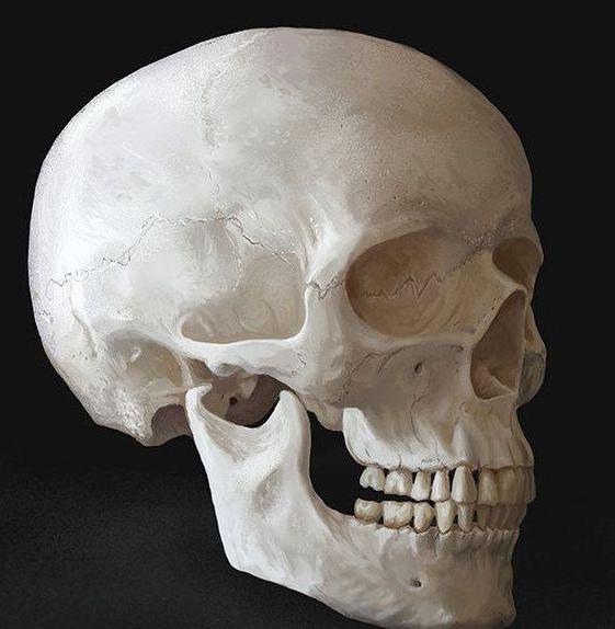
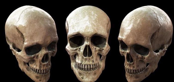
There are several criteria by which a male skull differs from a female skull. Such signs include the severity of roughness and tuberosity in the areas of muscle attachment, development of the mastoid processes and the occipital external tubercle, protrusions of the upper jaw, etc. The male skull is more developed than the female skull. Its outlines are more angular due to the severity of roughness and tuberosity in the areas of attachment of the masticatory, temporal, occipital and cervical muscles. Frontal and parietal tubercles are more developed in women, in men - epiglottis and superciliary arches. the latter have a heavier and larger lower jaw. In the area of the lower edge and the corners of the inner part of the chin pronounced tuberosity. This is due to the attachment of biceps, chewing and pterygoid muscles. Depending on gender, the shape of the human skull also varies. In men, a sloping forehead is noted, which turns into a rounded crown. Often there is a hill in the direction of the swept seam. Women's forehead is more vertical. He goes into a flat crown. In men, the orbits are lower. As a rule, they have a rectangular shape. Their upper edge is thickened. In women, the orbits are located higher. They are close to oval or round with upper sharper and thinner edges. On the female skull, the alveolar process in the upper jaw often comes forward. The nose-palate angle in men is pronounced in most cases distinctly. On the female skull, the frontal bone to the nasal passes more smoothly.

Additionally
The shape of the human skull does not affect mental abilities. According to the results of numerous studies by anthropologists, it can be concluded that there is no reason to believe that the size of the brain region predominates in any race. Bushmen, pygmies, and some other tribes have slightly smaller head sizes than other people. This is due to their small growth. Often, a decrease in the size of the head can be the result of poor nutrition for centuries and the influence of other adverse factors.