Saliva plays a very important role in the human body. With its help, chewing food is glued together, swallowed, as well as taste perception and protection of tooth enamel from damage. And special glands secrete saliva, which will be discussed later.
Types of organs producing saliva
Excretory ducts of the salivary glands fall into the oral cavity, which are divided into large (have the structure of an organ) and small, which are located in different parts of the mucosa.
Small ones include labial, buccal, molar, lingual, and palatine. Big are called two parotid, submandibular and sublingual. The largest is a pair of parotid glands.
Physiology
The salivary glands during salivation release a secret through the duct system into the oral cavity, which contains enzymes involved in digestion: amylase, proteinase, lipase, etc. The secret of all the organs that produce them is mixed in the human mouth and form saliva, forming a food lump and providing the beginning digestion process.
Parotid glands
These two glands are considered the most important. They lie around the jaw branch and take part in the initial phase of digestion, secreting the necessary amount of secretion. They are of the serous type and produce ptyalin. Their secretions enter the oral cavity through the ducts of the parotid salivary glands.
These organs are located behind the branches of the lower jaw and in front of the mastoid process extending from the bone of the temple. They are closely related to the functioning of the branching of the facial nerve, therefore, if their functioning is disturbed, serious dysfunction in the movement of the facial muscles can occur.
Through the excretory ducts of the parotid salivary glands , almost a fifth of the total volume of saliva enters the oral cavity. The weight of each of them ranges from 20-30 g.
Submandibular gland
The submandibular salivary glands produce a mixture of mucus and serous fluid. Despite the fact that they are smaller than the parotid, the proportion of salivary fluid produced by them is 70%. It enters the oral cavity from these secretory organs with the help of the submandibular canal, which is the duct for these salivary glands.
Description of the hyoid gland
Sublingual or sublingual are called large, under the tongue, glands. They are mainly involved in the secretion of mucus. Unlike other large glands, the duct system of the hyoid salivary gland is simpler. She is not so diverse and branched. It does not include plug-in ducts and jet flow outlets.
From the sublingual glands, salivary ducts in an amount of 8 to 20 open into the oral cavity. Up to 5% of all saliva passes through them.
The structure of the parotid glands
The parotid glands are a complex alveolar gland. Each of them has a lobed structure and is covered with fascia, which closes them into a separate capsule formation.
The excretory duct of the parotid salivary gland opens into the oral cavity in the form of a small hole located in front of the second large molar in the upper jaw. Its length is 6 cm and on the way to the oral cavity it passes through the surface of the masticatory muscle, adipose tissue of the cheek and buccal muscle. Sometimes this duct can bifurcate.
The structure of the submandibular gland
In its anatomy, it acts as a complex alveolar-tubular gland, the second largest in size among large organs that secrete saliva. It, like the parotid, has a lobed structure and is located in the submandibular fossa, extending beyond the posterior border of the maxillary hyoid muscle. The base of the salivary gland duct, located under the jaw, is located near the posterior edge of this muscle and, bending around its surface, opens on the hyoid papilla.
The structure of the hyoid gland
The structure of this gland is the same as that of the submandibular. It is located immediately below the oral mucosa over the maxillary hyoid muscle. There, it forms a sublingual fold located between the surface of the lower jaw and the tongue. The number of ducts of this gland can vary from 18 to 20. They open into the oral cavity along the hyoid fold. The main duct of the salivary gland passes near the submandibular ducts and opens with it by a common opening or near it.
Functions
The main purpose of the described glands is to develop a special secret. The ducts of the salivary glands are intended for its excretion of the oral cavity. Thus, the functioning of the salivary ducts provides the following:
- the oral cavity is wetted with saliva;
- food liquefies;
- articulation is provided;
- increased taste sensations;
- teeth are protected from damage (thermal or mechanical);
- the oral cavity is cleaned.
Possible diseases
There are many diseases that can disrupt the functioning of the salivary glands and the ducts branching from them. Among them, the most dangerous are:
- Duct expansion. It leads to a violation of the secretion in the oral cavity and becomes the cause of the formation of stones and purulent inflammation in the ducts of the salivary glands.
- Abscesses. This disease affects the glandular tissue, therefore, requires urgent hospitalization with subsequent surgery.
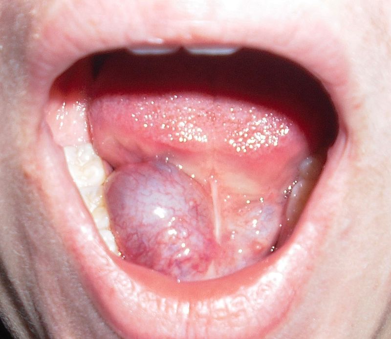
- The formation of intraglandular stones. In the process of the development of the disease, the system of ducts of the salivary glands is filled with stones, which complicate the passage of the secret.
- Sialadenitis. With the onset of the disease, there is a decrease in the secretion activity by the gland, leading to inflammatory processes that spread in the gland itself and its ducts.
- The formation of polyps that block the movement of the secret. As a result of constant fluid stagnation, the development of infection and inflammation begins.
- Sialolithiasis. The course of the disease involves filling the ducts of the glands with stones, leading to the same consequences as polyps.
- Mukocele. There is stagnation of saliva accumulated in the ducts arising due to polyps or stones.
- Papillary stenosis. Due to the disease, the ducts of the salivary glands narrow in places where the secret goes into the oral cavity, which leads to its stagnation and the development of the inflammatory process.
Treatment methods
In the vast majority of cases, diseases affecting the salivary glands and their ducts are treated by surgical intervention. The reason is that patients rarely seek help in the early stages of the development of the disease, and since delaying treatment leads to complications of the disease, only a surgeon can get rid of them.
Surgical treatment includes the following measures:
- Lithotripsy. During this procedure, the doctor using a special apparatus crushes stones in the salivary gland or duct and then extracts them.
- Marsupialization of ducts. Treatment is performed by opening the salivary duct, from which stones or polyps are extracted. Since more gentle methods currently exist, marsupialization is used very rarely and only in cases where large stones or a formation at the bottom of the mouth are found. After extracting the pathological formation, plastic ducts are made.
- Therapeutic sialoendoscopy. It is a variant of endoscopic surgery and makes it possible to remove the formed stones of a small size, and also get rid of strictures (narrowing of the lumen) of the ducts. A procedure is performed under local anesthesia by introducing a tube (or several) into the duct.
- Extracorporeal lithotripsy. The impact on the stones formed in the duct from the outside with the help of a special emitter is provided. In the process of such treatment, stones are destroyed regardless of their size. After crushing, the stones are removed and the ducts are washed with a special solution to prevent the development of inflammation.
- Endoscopic laser lithotripsy. This method is based on the direct effect on the stones in the duct. Crushing is carried out using a laser emitter. At the end of the procedure, the stones are removed.
- Endoscopic removal of polyps. The procedure is performed using a laser, which cut off the polyps. It is very popular because the laser, after cutting off the polyp, cauterizes and disinfects the place where the growth was located. In addition, there is no bleeding of the ducts of the salivary glands, which prevents the development of purulent complications.
- Endoscopic dilatation. It is used in cases of need to dissect adhesions in the gland or duct, which are formed on the scar tissue during the disease of the salivary glands. The procedure allows you to restore the outflow of secretion without damaging the walls of the ducts.
Endoscopic methods of treating diseases affecting the salivary glands and ducts are very popular, as they are highly effective and do not require further hospitalization. In addition, they prevent the development of various complications, which allows patients to recover quickly.
Since the salivary ducts play a very important role in the process of salivation, any disturbance in their functioning leads to serious consequences. Therefore, at the first sensation of discomfort in the salivary system, you must consult a doctor who can make the correct diagnosis and prescribe an effective treatment method.