Cattle disease is an important topic in modern veterinary medicine. Conventionally, all pathologies are divided into infectious and not having such a nature. The most dangerous are diseases that belong to the first class, especially those that can spread to a person. Infection of at least one animal is associated with the risk of losing an impressive percentage of the total population. If the disease is not contagious, it is relatively safe for others, but can be fatal. Suspecting an individual's disease, it is necessary to urgently consult a veterinarian and take treatment measures.
Infections
Such pathologies can appear against the background of the penetration of a virulent virus, pathological bacteria, and fungus into the body. Infectious diseases of cattle are dangerous infectious, can spread easily inside the herd. Infection of only one animal can cause the disease of the entire livestock that the farmer has - and this entails serious financial losses. Most often, infection leads to foot and mouth disease and smallpox. Cows can also get tuberculosis, brucellosis. Infectious diseases include rabies, actinomycosis, leukemia.
Brucellosis
The name cattle infectious disease was given by the pathogen - brucella. This bacterium is dangerous for animals; it can be transmitted to humans. The reproductive organs are the first to suffer, and infection occurs if healthy and sick cattle come into contact, graze in the same territory, and drink from the same source. The causative agent is able to penetrate the body through microscopic injuries of the skin, reproductive and respiratory organs, and through the digestive tract.
The disease does not manifest itself immediately, the latent period reaches three weeks. Brucellosis may be indicated by spontaneous interruption of the bearing of the calf, retention of the placenta, inflammation of the udder, endometritis. In bulls testicles, prepuce are inflamed. Regardless of the sex of the animal, brucellosis can cause abscesses and joint diseases.
The therapeutic course involves the appointment of broad-spectrum antimicrobial agents. As a rule, medicines are chosen for levofloxacin, which quickly inhibit the vital activity of the pathogen. The veterinarian usually prescribes Lexoflon for five days or more. Until the program is completed, and the recovery is not confirmed, you can not use either milk or meat of a sick animal. After termination of the course, the individual is double checked for infection. Permission to use the product is given if both analyzes give a negative result.
Rabies
This is the name for acute cattle viral disease, which violates the functionality of the nervous system. The primary outcome is death. Wild and living animals can become ill. All kinds are subject to this ailment. More often young individuals become infected. The disease can be transmitted to humans. A distinctive feature of the pathogen is thermolability. When the medium is heated to 60 degrees, destruction occurs in 5-10 minutes. At the same time, the pathogen is resistant to low temperatures. Under the influence of acidic environments, alkalis inactivated, shows resistance to iodine, phenol.
When rabies is considered among other diseases of cattle in veterinary medicine, a high level of contagiousness must be paid attention to. Infection usually comes from an infected individual: the virus can spread with saliva. Often, transmission occurs with a bite. In a substance, microflora can exist up to 10 days. From the point of penetration into the body, the pathogen enters the brain through the trunks of nerves, infects NS cells. The latent period lasts a day or more, occasionally - a month or more. More often, the first symptoms can be noticed approximately 3-6 weeks after infection.
Features of the disease
Usually rabies in animals is a cattle disease that proceeds in a quiet form. The sick individual hoarsely hoarsely, saliva is actively released from her. The animal is shaky, limbs paralyzed, appetite changes. If the pathology develops violently, the cows become aggressive, which becomes especially noticeable when dogs approach them. A sick animal is trying to break loose, can throw itself at walls, roar hoarsely. Some tend to dig hooves in the ground.
To diagnose the disease, it is necessary to evaluate the clinical manifestations, epizootological signs. For this disease, cattle treatment is not provided. The infected individual is isolated, the doctor is called. After death, an autopsy confirms infection with the causative agent of rabies. Prevention of cases involves the timely administration of vaccines. Keep livestock away from stray dogs. An animal that has bitten a person must be isolated and observed for at least 30 days.
Foot and mouth disease
This term refers to a pathological condition provoked by an RNA-containing virus. Foot and mouth disease occurs when infected with an aphthovirus from the Picornaviride family. After penetration into organic tissues, the pathogen accumulates in the epithelial cells, which leads to the formation of aphthous foci. When diagnosing cattle disease, it is necessary to pay attention to the presence of vesicles filled with a specific fluid. By the time the maximum possible concentration is reached, the pathogen gets a chance to enter the circulatory system, which provokes a sharp deterioration in the condition of the individual.
Foot and mouth disease can be suspected by an increase in temperature (up to 41.5 degrees), loss of appetite, abundant foamy saliva and smacking. Aphthae appear on the oral mucosa. They can be seen in the area of the udder, the gap of the hooves, near the scrotum in the male animal.
There are benign and malignant forms of cattle disease. The first option involves the absence of a secondary infection, and a complete cure is possible in two weeks. If the pathology is complicated, it will not work to avoid a fatal outcome. The greatest risks are for small calves aged three months and younger. Symptoms in young animals are more like gastroenteritis than foot-and-mouth disease, and pathology does not lead to the formation of aphthae.
How to get rid
When cattle diseases appear, their symptoms and signs must necessarily cause anxiety in the owner of the animal. You must call a veterinarian to determine the exact diagnosis. When confirming foot-and-mouth disease, a special serum should be used, due to which the body's ability to resist the harmful virus increases. Aphthae are regularly treated with furacilin, potassium permanganate, synthomycin ointment. To prevent re-infection, it is reasonable to prescribe an antimicrobial course. Drugs are given in liquid form along with food. If a sick individual refuses to eat, a flour mill should be introduced directly into the digestive tract through a probe.
Parainfluenza
This cattle disease also belongs to the class of viral, belongs to the group of contagious. The respiratory system is the first to suffer, it is here that the main disorders associated with the introduction of the pathogen are localized. More often the disease is observed in young livestock - no younger than ten days old, but no older than six months old. The disease provokes paramyxovirus, characterized by poor resistance to disinfectants. For inactivation, you can use alkaline, acid solutions, ether, chloroform.
Usually the infection comes from a virus carrier or a sick individual. Transmission - by airborne droplets. A sick cow is thought to be able to infect a calf through milk. There is no accurate information about the impossibility of sexual spread of the virus. More often, pathology is observed in cold weather, against the background of severe stresses, crowded habitat and the need for transportation.
Pathology Features
Parainfluenza is a common disease of young cattle, so it has been studied quite well. It was revealed that the incubation period usually lasts 24-30 hours, after which the symptoms of the disease are immediately noticeable: nasal mucous membranes turn red, tears are released, flowing from the nose, breathing is disturbed. A sick animal is depressed, body temperature is increased, inflammation of the ocular cornea, cough is observed.
To clarify the diagnosis, it is necessary to take a smear, swabs for laboratory analysis. Samples are obtained between the second and fifth days of the disease. It is necessary to examine blood plasma in the first three days. Symptoms of parainfluenza are similar to other respiratory diseases caused by viruses, so special care is required.
Having identified the disease in young cattle, it is necessary to immediately isolate the individual. This animal has been treated, among other things - vaccination. The therapeutic course involves the introduction of special serum, globulin, convalescents. Antimicrobials are used, funds from the group of nitrofurans, sulfonamides.
Prevention of parainfluenza is possible if you carefully observe sanitary, veterinary, technological measures, take care of livestock and keep it in good conditions, and regularly vaccinate young livestock. An ill animal in the future cannot be re-infected. Newborn animals with colostrum receive antibodies when fed if the cow has been vaccinated. It is recommended to vaccinate animals on the 5-7th day of birth, when the antibodies received from the mother cease to act.
Smallpox
This is a cattle disease provoked by several pathogens. Possible cow, pork and smallpox vaccines. A distinctive feature is the formation on the outer covers of clearly defined papules, the central part of which is somewhat depressed. Diseases are characterized by an acute course, fever, general poisoning of the body. Smallpox can be suspected if the animal is not eating, weak, rashes of pinkish papules are formed on its lips, udder, near the nose, gradually changing color to darker. Formations gradually burst, exudate flows out, crusts appear. The animal often lies, and if it gets up, it widely spreads its legs when walking. Measurement of body temperature shows elevated rates. Often, the pathology is complicated by mastitis. To avoid this, milk is decanted regularly. If it is impossible to do this with your hands, you need to use a catheter.
The therapeutic course involves the treatment of all rashes with formalin or brilliant green. Stimulation of regeneration is possible when using boric or zinc ointment. Improving the immune status is provided by vitamin supplements to the main diet. To prevent secondary bacterial infection, antibiotics are prescribed for streptomycin, bicillin.
To prevent smallpox, it is necessary to plant livestock on time. This is especially important if there have already been cases of diseases in the area.
Tendon diseases and bruises
In cattle, diseases of the joints and tendons are frequent, as well as a variety of injuries associated with a lifestyle. Many have tendonitis, tendovaginitis. Such are especially frequent when an individual receives injuries, wounds, as well as against the background of infection. The sick area becomes thicker, swollen, sore. The animal is limping, when touched, fever is noticeable. The therapeutic course involves ensuring complete rest, applying a pressure dressing, cooling the site. If excess effusion accumulates, it is necessary to make punctures, treating the area with antiseptic solutions. In this case, wet compresses with ichthyol alcohol, camphor should be used. When the pain subsides, camphor oil is used for local massage.
Among cattle limb diseases, bruises are very common. Of course, the animal can get such an injury in any part of the body, but it is the legs that are more likely to suffer. A bruise is a mechanical damage in which the skin remains intact. Bruising is possible by contact with the hoof, mechanism, or a blunt object. The animal can receive this kind of damage during the transportation period. If the bruise is severe, nearby tissues may become inflamed. In addition, there is a risk of fracture. The affected area is hot to the touch, responds with pain, after a few days, discoloration of the skin is noticeable. At first they become bluish-black, and then yellowish-green. If the wound is deep, the animal may have a febrile condition that is not accompanied by infection.
The affected individual needs rest. The skin is treated with iodine, potassium permanganate. In case of severe damage, it is reasonable to use cold, apply a compress or pressure dressing. The treatment is chosen by the veterinarian, focusing on the condition of the animal. Bruises can be prevented if livestock are kept in adequate conditions, dividing by sex and age, observing the basic rules of transportation.
Livestock pathologies: different parts of the body suffer
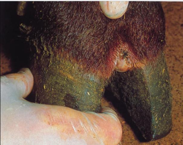
Cattle hoof diseases are quite common in cattle. All of them bring animals a lot of problems and anxieties, since walking becomes a source of pain, as well as being in a standing position. Quite often, the so-called strawberry disease develops, in which small reddish tubercles form at the base of the slit and on the diadem. With their infiltration, laminitis is diagnosed. Perhaps the appearance of a corolla phlegmon, in which the tissues become inflamed, purulent exudate is released.
To diseases of hooves in cattle include lameness. The term denotes a condition accompanied by active growth of the stratum corneum. This is more typical of the period of prolonged stay in the stall. In inflammatory processes at the base of the hoof, aseptic pododermatitis is diagnosed.
To identify the problem, it is enough to carefully monitor the behavior of the animal. A sick individual tends to lie, limps when walking. A visual examination shows swelling and enlargement of the joints, deformation of individual areas. Perhaps the formation of foci of ulceration, abscesses, purulent substance is secreted from the crack of the hoof. With these symptoms, you should not look for information with photos about cattle diseases in directories and other sources. A veterinarian should be invited so that the specialist makes an accurate diagnosis and selects the appropriate treatment.
Tuberculosis
Invasive cattle diseases include tuberculosis. Provokes the disease Koch's wand. When it penetrates into organic tissues, a starting focus is first formed, from where further spread gradually occurs with the generation of multiple lesion zones. Gradually, organic tissues in the implantation area are destroyed. Forms vary from case to case, depending on the location of the pathogen. It is customary to talk about the pathology of the bone system, respiratory, intestinal tract and uterus. There is the likelihood of a generalized option, in which the wand penetrates the circulatory system and simultaneously infects several organs.
The disease is transmitted not only between livestock within the herd, but can infect humans. Symptoms are far from always expressed, in adult animals it can proceed secretly. Most often, the area of localization is the lungs. A sick individual loses appetite, is exhausted, suffers from shortness of breath. A fever is possible, but quite slight, as well as a cough. Lymph nodes become larger in size, lose mobility, and listening to the lungs allows you to determine wheezing.
Other forms of tuberculosis
A different area of localization of the Koch bacillus gives other manifestations. So, with infection of the udder, the back and the lymph node above the organ grows. With intestinal, loose stools with inclusions of pus and blood are noticeable. The proliferation of lymph nodes in different parts of the body that respond with pain can indicate a generalized form.
To confirm the diagnosis, it is necessary to put a tuberculin test. Normally, they do it to the whole herd. Veterinary recommendations contain an indication of the timing of the injection. When determining a positive response, an individual is subject to slaughter. When an infected animal is detected, the economy goes into the category of dysfunctional. It is necessary to completely replace the herd, to disinfect all objects related to the keeping of animals.
Telaziosis
Pathology is provoked by telazii, infecting the lacrimal glands, other parts of the eye. The intermediate owner of the nematode is the barn. Typically, infection with cattle eye disease occurs during the walking period on pastures. The fly feeds on the secretions of the animal, while swallowing the larvae, which then accumulate in the head and through the proboscis move into the eye of the mammal. The duration of this phase of the life cycle reaches 11 months. . , . , , . . . .
To clarify the diagnosis, it is necessary to take a flush of the substance obtained from the conjunctival sac for examination. Telasiosis is diagnosed with the identification of larvae, adult parasites. Noticing the symptoms of the disease in cattle, it is shown to do deworming. With complications of secondary infection, sulfonamides and penicillin antibiotics are indicated. Deworming involves the use of a half-percent iodine solution, an ichthyol emulsion, lysol made from fish fat, and a solution of boric acid. The drug is administered under the third eyelid in an amount of not more than three milliliters, after which the individual's eyes are massaged. It is necessary to do three such procedures, having passed between them an interval of no more than three days.
In order to minimize the risks of the disease, regular preventive deworming should be carried out and flies should be controlled. Prevention of infection involves the implementation of preventive measures before the start of grazing, while cattle are kept in stalls.
Hepatitis
Among non-communicable diseases of cattle, hepatitis is one of the most common. The term refers to diffuse inflammatory processes that occur in the liver. Pathology leads to hepatic hyperemia, tissue infiltration. Dystrophic processes, necrotic changes are initiated. Liver cells and other structural elements associated with them are affected. The disease manifests itself as signs of liver failure. Hepatitis usually occurs if an individual eats spoiled food, lupine, and potato sprouts. Similar symptoms are possible with almond poisons. Some invasive pathologies can lead to hepatitis.
Hepatitis is a non-infectious disease of cattle, which manifests itself in a decrease in appetite and general inhibition of a sick individual. The animal is thirsty, he begins to vomit, fever develops, breathing becomes frequent, secretions with bloody inclusions are plentifully secreted from the nose, the mucous membranes acquire a yellowish tint, the skin patches itch, the animal combes the area to the blood, and urine acquires a dark shade. The duration of the acute period reaches a month, after which the livestock recovers or dies. With the transition to a chronic form, hepatic cirrhosis begins, the organ becomes denser, functions weaken. A blood test shows a high concentration of bilirubin.
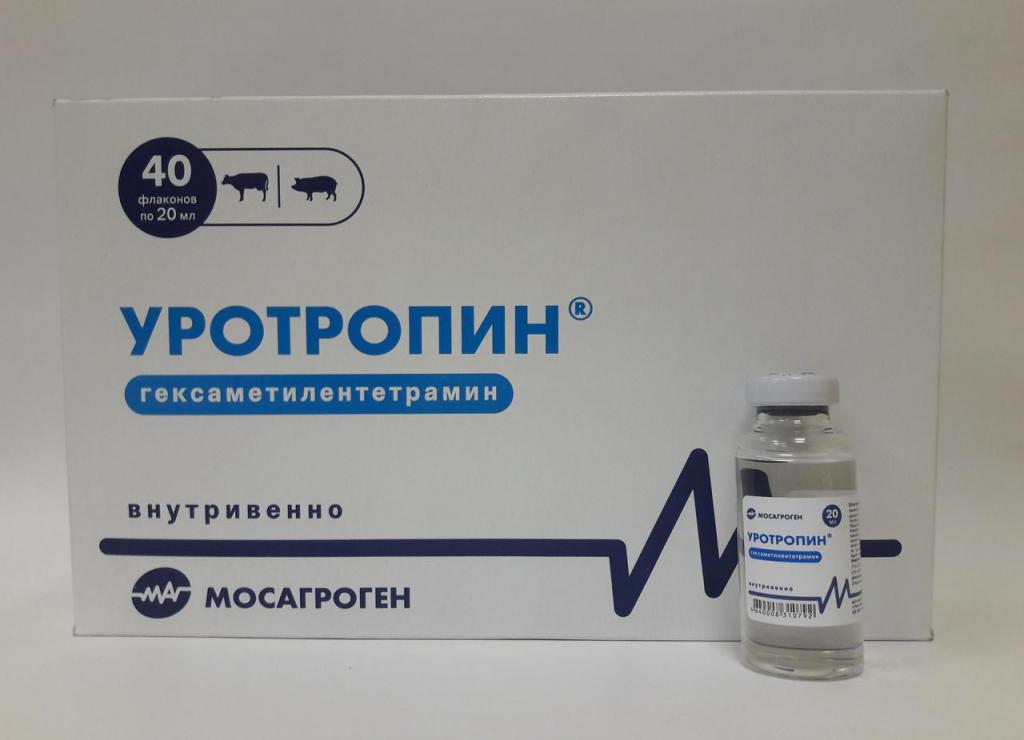
If this internal non-communicable disease of cattle is detected, it is necessary to transfer to a diet. Coarse forage crops, carbohydrates are shown. A glucose solution must be injected into a vein, and magnesium sulfate orally. The therapeutic course involves the use of "Urotropin", Karlovy Vary salt.
Plague
The long-known acute viral disease that scares many farmers and affects livestock is the plague. Infection proceeds systemically, provokes a severe febrile state, catarrhal and hemorrhagic manifestations. On the mucous membranes, you can see areas of inflammation. The likelihood of death varies between 90-100%.
This cattle disease history is quite long. For the first time the term "plague" was used in 1710. It is formed from the Greek word for general infection. Then the plague, it was decided to call any disease that caused significant damage. Back in the period of the Roman Empire, outbreaks of cattle plague were recorded. In Asia, officially for the first time this happened only in the fourth century. In European countries, the disease was especially widespread in the eighteenth century, as a consequence of hostilities and active trade between countries. First, German and Dutch, English and Italian lands suffered, then the pathology spread to the Scandinavian powers. There was a place for this cattle disease in the history of almost all countries in Eurasia.
Quite strong outbreaks were observed until the third decade of the last century. The damage from them can not be estimated, it is so great. In the period of the 60-80s of the nineteenth century, about 200 million individuals died in Europe alone. Outbreaks of the disease were regularly observed in Asia and the Far East.
Propagation and Frequency
In our century, most often the plague appears in African and Asian states. Such cases are noted in powers where the maintenance of the virus is ensured by a population of wild fauna. According to information collected by the OIE, in 1976-1980 about 15 powers in Africa remained extremely disadvantaged on the issue of the plague, where outbreaks were noted every year. Most often this happened in Sudan.
Among Asian countries, plague is prevalent in 12 countries, of which eight are in the Middle East. The most pressing issue for India and Kuwait.
To study the course of the disease, healthy individuals were infected with the plague pathogen. Tests have shown: the duration of the latent period reaches a week. With natural infection, the interval varies from three days to two and a half weeks. The course is usually acute, somewhat less often - subacute, super acute. As a rule, at first there is a fever, the fever persists for several days, in the morning some relief is possible. A sick individual makes a grinding of teeth, ruffles the hair. The mirror of the nose is dry, foci of inflammation are visible on the mucous membranes of the eyes, nose, and mouth. You can see separate areas of redness, often near the gums, nodules of a gray and yellowish tint are gradually formed. The epithelium dies, forming a yellow cyst with a specific smell, ulcers appearing with this are characterized by uneven edges. Saliva liberates abundantly in the animal, eyes, nasal mucous membranes become inflamed, vaginitis is observed. First, the character is serous, purulent discharge gradually appears.
Under stably unfavorable conditions, benign forms are more often observed due to residual immunity. Symptoms are rather mild, on the mucous membranes there are usually no foci of necrosis, there is a chance of a full recovery. Fatal cases mainly occur in young animals, their frequency does not exceed 40%.