The underground organ of most higher spore, gymnosperms and flowering plants is the root. For the first time, it appears among the plunders and performs not only the function of support, but also provides all other parts of the plant with water and mineral salts dissolved in it. In gymnosperms and angiosperms, the main root develops from the germinal root. Subsequently, the root system is formed, the structure of which differs in monocotyledonous and dicotyledonous plants. In our article, we will study the primary and secondary anatomical structure of the root of flowering plants, the seeds of which have two cotyledons, and using concrete examples we show the role of plant tissues and structural elements of the underground part in ensuring the vital functions of the plant organism.
Germ root and its development
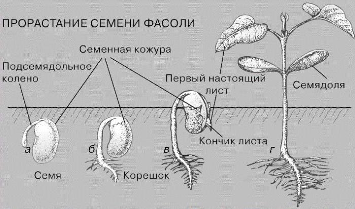
In the process of seed germination, the first part of the embryo, called the germinal root, develops. It consists of cells of the educational tissue - the primary meristem, the apical part of which is called the apex. In the process of mitotic division of its constituent cells, the primary structure of the root is formed, consisting of an epiblem, a primary cortex and an axial cylinder. Let us dwell on the morphological and physiological characteristics of the primary educational tissue located at the apex of both the germinal root and the apical part of all young roots: the main, lateral, and adnexal. The species called the latter is found mainly in monocotyledonous plants. They develop from the bottom of the stem. So, the apex consists of initial cells. In the process of development, they form the primary meristem. Under its layer, the differentiation of cellular structures begins, leading to the appearance of a formed educational tissue, which determines the primary anatomical structure of the root. In a plant, it persists until the appearance of secondary meristems called cambium and pellogen.
Epiblema: structure and significance
Rhizoderma, or epiblema, is a layer of integumentary tissue cells located on the young central root and lateral processes extending from it. The most important for the plant is the part of the integumentary tissue, which is located in the root zone, which absorbs water and mineral salts. In it, elongated epiblema cells form root hairs. Their cytoplasm contains a large number of vacuoles, and the cell wall is very thin, without cuticle. The rhizoderm is located on the root site from the root cap to the zone of lateral roots, which is called conductive. It has been established that the position of root hairs with respect to the root cap located at the apex of the main root practically does not change.
Root hairs and their role in plant life
Examining under the microscope the primary structure of the root, it can be found that the rhizoderm is a derivative of the uppermost layer - the dermatogen. It, in turn, is formed as a result of cell division of the primary apex. The suction zone of the root is most sensitive to sudden changes in environmental conditions, therefore, measles hairs can quickly die off. This is the main reason for poor survival of seedlings and even its death. In the process of seedling development, the rhizoderm cells die and slough. Under them, a layer of protective tissue is formed - the exoderm, which partially takes part in the formation of access elements. Thanks to them, water and solutions of mineral compounds from root hairs enter the axial cylinder, which is part of the primary structure of the root.
It contains conductive tissues, from which vessels develop during the process of ontogenesis - trachea and sieve tubes with companion cells. Not all plants form a developed system of root hairs. For example, in wetland and aquatic species they are absent due to excess water in the environment.
Primary Meristem - Pericycle
This is a structure that, in the form of a ring, covers the central cylinder and is located under the rhizoderm. It is represented by small, rapidly dividing cells of the educational tissue and is present in all woody and herbaceous plant forms propagated by seeds. All parts of the central cylinder develop precisely from the cells of the pericycle.
The primary structure of the root of the dicotyledonous plant confirms the fact of laying the lateral and adnexal roots in the outer layer of the educational tissue - the meristem. In representatives of dicotyledonous plants belonging to the families of Rosaceae, Legumes, Solanaceae, it is then converted into secondary species, for example, phallogen or cambium. The result of mitotic cell division of the pericycle is the appearance of the germinal zones of future tissues that are homogeneous in structure and function - the periblema from which the primary cortex is formed, and the dermatogen, which gives rise to the apical primary meristem.
Primary bark
This part of the root is represented mainly by parenchyma cells. The part of the plant tissue adjacent to the epiblema is called exoderm, the middle layer of the primary cortex is called mesoderm. Examining the primary root structure under a microscope, a large number of intercellular spaces can be found in these areas. They serve as a place of circulation of oxygen and carbon dioxide, which means that they participate in gas exchange. The inner portion is represented by groups of cells arranged in the form of a dense strand.
After the destruction of the epiblema, the exoderm sections are exposed, then they are sampled in the zone of lateral roots and subsequently perform a protective function. Through all three layers of the cortex, water molecules move in the radial direction and then enter the vessels of the central root cylinder. According to them, due to root pressure and transpiration, water and solutions of mineral substances rise into the stem and leaves. In addition, organic compounds such as starch or inulin can accumulate in the parenchymal cells of the mesoderm of the primary cortex.
Central cylinder
Examining under the microscope the primary structure of the root of a dicotyledonous plant, one can find such a structure as a stele. This axial part contains several anatomical formations that perform the functions of conducting substances. They consist of primary tissue - xylem and form conductive elements such as vessels (trachea). Solutions of glucose and other organic compounds move from leaves and stems to the root through sieve tubes located in the cortex, and water and mineral substances through the vessels (trachea) come from the root axial cylinder to the vegetative organs of the plant.
The role of cambium in root development
The transition from the primary structure of the root to the secondary occurs at the seedling stage and is marked by the appearance of educational tissue - cambium. One of its species is formed from the protomeristem of vascular bundles.
Then there are sections of radiation cambium. Both of these varieties of the secondary meristem merge into a common cambial ring lying between the cortex and the central cylinder. Due to active mitotic division, cambium cells form two layers of secondary conducting tissues: the inner, directed towards the stela - the xylem and the peripheral, facing the endoderm - the phloem. As a result of the above processes, the axial cylinder acquires a secondary structure characteristic of all the roots of dicotyledonous plants.
What changes are occurring in the primary cortex
The appearance of secondary conductive tissues - phloem and xylem causes transformation in the pericycle. Its cells, dividing by mitosis, form a layer of cork cambium - the phallogen, which, in turn, forms the periderm. An integral part of its cells begins to divide periclinally, which leads to isolation of the primary cortex from the axial cylinder, and then to its death. Now the outer layer of the secondary root is the periderm with the remaining parts of the phalloderm and pericycle. As you can see, the primary and secondary structure of the root are radically different from each other. These differences concern all its departments, including the cortex and central cylinder. They are especially noticeable in the anatomical structure of educational and integumentary tissues. The most important processes occurring in the root during its growth can be considered the appearance of cambium and the laying of secondary conductive tissues. In the next subheading we will consider them in more detail.
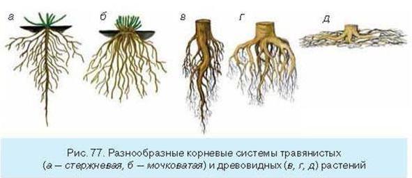
Primary and secondary root structure
Differences in the morphology and physiological functions of the growing root of a dicotyledonous plant can be represented in the form of a table:
| Germ root | Young plant root |
| Covering tissue (epiblema) | Coating tissue (capped exoderm) |
| Primary cortex: exoderm, mesoderm and endoderm | The secondary bark is formed by cambium (bast) |
| Stela: pericycle, primary xylem | Stela (secondary xylem) |
| Cambia no | Secondary Meristem (Cambium) |
In addition to the table, we note that the secondary thickening of the root of roots in dicotyledonous plants is explained by mitotic activity of cambium cells, and root growth in length is associated with the renewal and movement of apical meristem and root cap cells deep into the soil layer. The top of the central root overcomes the resistance of solid sections of the soil due to its high growth energy, which is why the roots of woody species of angiosperms during germination can penetrate even asphalt.