Many are interested in echocardiography with Doppler analysis and CDK. What is it, we will understand in this article.
The heart is the most necessary and, most importantly, irreplaceable part of the human body. It knows no peace and works tirelessly throughout human life, without stopping the movement of blood through the vessels. As applied to the heart, the phrase that movement is life is not an idiom, but a very real fact. Disorders in the work of the heart are the most relevant medical problems to date, attracting doctors to the choice of methods for identifying various pathologies and curing them. Widely used in modern medical practice, this type of diagnosis, as echocardiography with Doppler analysis. How is this study conducted, what is it, how to prepare for it, and what information can be extracted with it for diagnosis?
History
Pulse ultrasound in heart research was first used by scientists in Sweden in 1954. They developed a device with which it was possible to receive signals from the mitral valve and the left ventricle. Over the past time, the technology has been significantly improved and has found its place in cardiology practice. Currently, this method is called echocardiography with Doppler analysis, and its main advantage is the visibility and obtaining a complete picture in a short time, which increases the chances of seeing even the most minor pathology.
Echocardiography: what is it?
The use of high-frequency waves that the human ear is unable to perceive, or ultrasound, is the essence of the method of echocardiography. Through special sensors attached to the body, ultrasonic waves propagate in the tissues, change their frequency and amplitude of oscillation depending on the state in which the internal organs are. Then the waves return to the sensors and, converted into an electrical signal, are processed by the device. This creates a picture of the study of the state of the heart muscle from four sides. The screen shows a two-dimensional or even three-dimensional image. In medicine, echocardiography is a diagnostic method that allows you to identify cardiac pathologies of a different nature in a wide variety of patients. For example, an echocardiographic study with dopplerometry and color Doppler mapping (CDL), which is a combination of M-modes and 2D with dopplerometry, is indispensable for assessing mitral stenosis. The method has practically no contraindications, the studies are carried out in a short time and the result is formed just as quickly - all this allows the active use of echocardiography in medical research. What is remarkable about echocardiography with Doppler analysis?
Features and Benefits
This procedure can be done along with adults by children and pregnant women. Echocardiography is the most accurate method known today.
According to doctors, this method has many advantages:
- Non-invasiveness of the method.
- The doctor receives a large amount of information quite quickly and quickly.
- Affordable for the price.
- Existing data is easily reproduced.
By methods of echocardiography with Doppler, the doctor determines the direction of blood flow, measures the speed of blood movement. But the method also has disadvantages. One of them is that in order to obtain the most reliable results, the passage of the ultrasound beam should be as parallel as possible to the blood flow. Of course, this limits the study of some parts of the heart.
Estimated Parameters
What parameters are evaluated by echocardiography with Doppler analysis and CDK?
Echocardiography data allows you to evaluate:
● valves and their functional features, as well as the structure of the heart, located next to the valves;
● any abnormalities in the communication between the ventricles;
● blood leakage when the valve is closed;
● volume of pumped blood.
Doppler echocardiography allows you to measure all of the above indicators, which are important for evaluating childhood cardiac pathologies and congenital malformations without the use of cardiac catheterization. In addition, echocardiography with Doppler analysis does not pose a danger to the baby, but can not be said about computed tomography, where the patient is exposed to x-ray radiation.
With the help of echocardiography, it is possible not only to obtain information about the heart muscle and blood vessels, but also to carry out studies of various kinds of tumors, diagnose mitral stenosis, and conduct many other examinations. How is echocardiography with Doppler analysis performed in newborns? About it further.
Who can be assigned?
This analysis for children is carried out in the same way as for adults. The procedure can be performed in people of any age. Moreover, it is prescribed for pregnant women. With echocardiography there are no unpleasant sensations, the procedure does not harm the child, makes it possible to learn about the location and structure of the vessels and heart of the child.
In what cases recommend echocardiography with Doppler analysis and CDK?
Indications for echocardiography (Echocardiography)
This procedure is examined by patients who underwent heart surgery. In addition, prolonged headaches can also be an indication for examination, since the origin of the pain may have a more serious reason than it might seem at first glance. Echocardiography is often prescribed for young children who are poorly gaining weight.
Indications for echocardiography are:
- deviations from the norm identified in previous studies of the chest;
- heart rhythm disturbance;
- heart murmur;
- pain in the chest area, which has unknown reasons;
- ischemia, acquired and congenital malformations, arterial hypertension, myocardial infarction.
Mandatory echocardiography with Doppler analysis and stress Echocardiography is prescribed for pregnant women if:
- the birth of children with congenital heart defects was observed;
- previous pregnancies were interrupted by a miscarriage;
- diagnosed with diabetes;
- rubella was found in a pregnant woman;
- were prescribed for taking antibiotics.
Echocardiography for pregnant women is prescribed for a period of 20-23 weeks, children can undergo it at any age. How is echocardiography with Doppler analysis performed?
Doppler echocardiography
The basis of this study is the use of ultrasound. Preparation for the study does not take much time and does not present any difficulties. Doppler echocardiography is often prescribed for a child. The patient strips to the waist, then lays on the couch. It is necessary to lie on your left side so that the chest and the top of the heart are closer to each other, this provides more accurate results. Then the doctor applies a gel to the chest where the sensors will be attached. They do not cause pain or discomfort. During the procedure, the doctor is facing the patient or behind his back. Ultrasound from sensors attached to a person is transmitted back to the body, then it will be converted into an electrical signal that the device processes. This is the difference between echocardiography and ECG. The results of the study in the form of a clear picture appear on the monitor with echocardiography with Doppler analysis. What is it, we explained.
Doppler options
With an echocardiogram, different doppler options are used:
- Pulse wave. Allows specialists to study the flow of blood in a particular vessel, in the area of a particular valve.
- Energetic. This option is used to register low-speed blood flow. To date, energy doppler is not yet widely used.
- Continuous wave. This Doppler variant is used to record high-speed blood flow.
- Tissue. Used to assess the speed of movement of the myocardium and other cardiac structures.
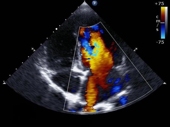
- Color. It is used for rapid assessment of blood flow in the main vessels, ventricles and atria of the heart.
- Color M-mode. In this case, a combination of the usual M-mode is used, which makes it possible to obtain a graphic image of the movement of the valve flaps, heart walls in time and the blood coding color mode.
Echocardiography with color mapping and Doppler analysis has become increasingly popular.
Why is dopplerography echocardiography performed?
Evaluation of the work of the heart muscle is impossible without studying the strength and volume of blood flow in the vessels and heart chambers. The use of modern ultrasound systems designed to perform echocardiography, equipped with dopplers and the necessary computer software, makes it possible to brilliantly solve this problem. Specialists have the ability to simultaneously conduct ultrasound scanning of the heart and Doppler ultrasound.
This diagnosis allows specialists to:
- identify pathological blood flow directions;
- to calculate the effective surface of the tricuspid foramen and mitral valve;
- determine what an indicator is like turbulence in blood flow;
- evaluate pressure indicators in the heart chambers, the amount of blood ejected by the heart in one reduction, diastolic compliance of the left ventricle, and other indicators of cardiohemodynamics.
Echocardiography in combination with Doppler allows specialists to obtain the necessary information to identify patients with heart defects and other pathologies. Echocardiography is most useful when the following pathologies are diagnosed:
● heart defects: with valve dysfunctions to control prostheses;
● dysfunctions of the left ventricle: used to find out the causes (post-infarction cardiosclerosis, cardiomyopathy, etc.) and determine the ejection fraction;
● atrial fibrillation - an assessment of the structural cause, the risk of thromboembolism, and the estimated response to cardioversion;
● chronic heart failure;
● cardiomyopathies;
● infectious endocarditis: valve lesions are assessed, as well as the severity of hemodynamic disturbances;
● conditions resulting from an ischemic stroke of the brain;
● pericardial pathology (presence of fluid in the pericardial sac);
● pathology of the thoracic aorta: aneurysm, dissection.
How to decipher the result of cardiac echocardiography with Doppler analysis?
Decoding Echocardiography
Cardiac echocardiography with Doppler and CDC may have contraindications if the patient was diagnosed with:
- acute myocardial infarction;
- heart failure;
- renal or liver failure;
- stratified aortic aneurysm.
Data from the study of the heart are deciphered as follows. First of all, a specialist evaluates myocardial contractility, and then checks the indicators of the left ventricle. A thorough check of its functional characteristics, size and condition of the cavity takes place, the presence of scars and various kinds of tumors, their sizes and the effect they have on the walls of the bloodstream are also examined. If the procedure was performed during physical activity, then it is believed that it passed normally in the presence of the following symptoms:
- the walls of the ventricle move evenly;
- an increase in the exile fraction is observed;
- the walls thicken.
It is believed that the study passed with a not very good result if:
- ejection fraction is less than 35%;
- the walls of the right ventricle increase in size;
- weak mobility of the walls of the ventricle is visible.
The results of the study should be analyzed by a cardiologist who compares normal values with those obtained in the study of this patient. Only on the basis of aggregate data, taking into account all circumstances, can a specialist make a diagnosis. Special preparation for echocardiography with Doppler analysis is not needed.
Cardiac echocardiography is of great importance, for example, in establishing diagnoses of atrial tumors, stenosis of the tricuspid valve. Thanks to this study, the doctor has the opportunity to compose a holistic picture of the disease based on important and very accurate diagnostic information.
Thus, echocardiography with Doppler analysis can be considered an extremely informative modern method for diagnosing cardiac pathologies. It allows doctors to check the structure of the heart, to track its work, as well as the movement of blood flow, its turbulence and speed. All information obtained during the course of the study is extremely important for determining the correct diagnosis. Nevertheless, it is always necessary to remember: a variety of factors, such as the type and quality of the equipment used, as well as the presence or absence of relevant experience from the doctor conducting the study, can influence the results of the study. An incorrect diagnosis due to an ultrasound scan usually leads to the appointment of unnecessary medical examinations, drugs and even surgical interventions.