The aorta is the largest vessel of the human body that carries blood from the left ventricle and is the beginning of a large circle of blood circulation.
In the aorta, several departments are distinguished:
- ascending (pars ascendens aortae) department;
- an arch and branches of an aortic arch;
- the descending (pars descendens aortae) section, which, in turn, is divided into the thoracic and abdominal parts.
Aortic arch and its branches
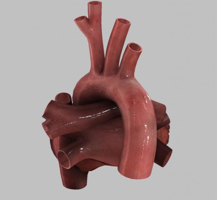
- Truncus brachiocephalicus branches off from the aortic arch at the level of the cartilage of the 2nd right rib. In front of it is the brachiocephalic right vein, and behind it is the trachea. After the departure, the brachiocephalic trunk goes up and to the right, giving in the region of the sternoclavicular right joint two branches: the subclavian right and the common carotid right artery.
- The common carotid artery (left) is one of the branches of the aortic arch. As a rule, this branch is 20-25 millimeters longer than the carotid common right artery. The path of the artery runs behind the scapular-hyoid and sternocleidomastoid muscle, then up the transverse processes of the cervical vertebrae. Outside of the vessel, the vagus nerve and jugular (internal) vein are located, inside of it lie the esophagus, trachea, pharynx, larynx, parathyroid and thyroid glands. In the area of the thyroid cartilage (its upper part), each of the common carotid arteries gives off the internal and external carotid arteries, which have approximately the same diameter. The site of the division of the artery is called bifurcation, in this place also lies an intersonal glomerulus (carotid gland, carotid gland) - an anatomical formation with dimensions of 1.5 x 2.5 mm, which is equipped with many chemoreceptors and a network of capillaries. In the area of discharge of the carotid external artery there is a small extension called the carotid sinus.
- The carotid external artery is one of the two terminal branches of the carotid common artery. It branches off from the latter in the region of the carotid triangle (the upper edge of the thyroid cartilage). Initially, it is located slightly medial to the carotid internal artery, and then laterally to it. The beginning of the carotid external artery lies under the sternocleidomastoid muscle, and in the region of the carotid triangle - under the subcutaneous muscle of the neck and cervical fascia (its surface plate). Situated inward from the biceps muscle (its posterior abdomen) and the stylohyoid muscle, the carotid (external) artery in the neck region of the mandibula (in the layer of the parotid gland) is divided into a pair of terminal branches: the maxillary and temporal superficial arteries. In addition, the carotid external atria gives rise to a number of branches along its course: the anterior group is the facial, thyroid superior and lingual arteries, the posterior group is the anterior posterior, occipital and sternocleidomastoid arteries, and the pharyngeal ascending artery goes to the middle.
Branches of the thoracic aorta
This segment, as already mentioned, is part of the descending aorta. It is located in the posterior mediastinum, passing along the spinal column.
The branches of the thoracic aorta are presented in two groups: parietal and visceral (visceral).
Intra branch
Visceral branches of the aorta are represented by the following groups:
- Branches are bronchial (2-4 pieces). Begin from the anterior wall of the aorta in the area of branching of the intercostal third arteries. Entering the gates of both lungs, they form the arterial intrabronchial network supplying the bronchi, connective tissue formations (frame) of the lungs, esophagus, pericardium, walls of the pulmonary vessels (veins and arteries). In lung tissue, bronchial branches form anastomoses with branches of the pulmonary arteries.
- Esophageal branches (3-4 pieces). They have a dyne of about 1.5 cm and end in the walls of the esophagus (its chest segment). These branches begin from the thoracic aorta in the region of 4-8 of the thoracic vertebra. Anastomoses are formed with the upper diaphragmatic, lower and upper thyroid, mediastinal arteries, as well as with the coronary left cardiac artery.
- Branches mediastinal (mediastinal) can have a variety of placement, intermittent. Often go as a part of pericardial branches. They provide blood supply to the fiber, lymph nodes of the posterior mediastinum and the wall of the (posterior) pericardium. Anastomoses form with the above branches.
- Pericardial branches (1-2 pieces) are thin and short. Branched from the anterior aortic wall, supplying the pericardium (its posterior wall). Anastomoses with mediastinal and esophageal arteries are formed.
Parietal branches
- The diaphragmatic superior arteries extending from the aorta circulate the pleura and the lumbar segment of the aorta. Combine into anastomoses with diaphragmatic lower, internal thoracic and intercostal lower arteries.
- The posterior intercostal arteries (10 pairs) branch off from the posterior aortic wall and follow at 3-11 intercostal spaces. The last pair passes under the 12th rib (i.e., is the hypochondrium) and enters the anastomosis with lumbar arterial branches. The first and second intercostal spaces are supplied with blood by the subclavian artery. The intercostal right arteries are slightly longer than the left ones and go under the pleura up to the costal angles, located posterior to the posterior mediastinum, lying on the front surfaces of the vertebral bodies. In the costal heads, dorsal branches extend from the intercostal arteries to the muscles and skin of the back, to the spinal cord (including its membranes) and the spine. From the costal angles, the arteries go between the internal and external intercostal muscles, lying in the costal groove. Arteries in the region of the 8th intercostal space and below it lie under the corresponding rib, branch into lateral branches to the muscles and skin of the lateral parts of the chest, and then form anastomoses with intercostal anterior branches from the thoracic (internal) artery. 4-6 intercostal arteries give branches to the mammary glands. The intercostal upper arteries supply the chest, and the three lower ones - the diaphragm and the abdominal wall (front). The third right intercostal artery gives a twig that goes to the right bronchus, and branches go from 1-5 intercostal arteries that supply the left bronchus. 3-6th intercostal arteries give rise to the esophagus arteries.
Branches of the abdominal aorta
The abdominal segment of the aorta is a continuation of its thoracic part. It starts from level 12 of the thoracic vertebra, passes through the aortic diaphragmatic opening and ends in the region of 4 vertebrae of the lower back.
The abdominal region is located in front of the
lumbar vertebrae, slightly to the left of the midline, lies retroperitoneally. To the right of it lies the
vena cava (inferior) vein, in front - the pancreas, a horizontal segment of the duodenum and mesenteric root of the small intestine.
Parietal branches
The following parietal branches of the abdominal aorta are distinguished:
- The lower diaphragmatic arteries (right and left) branch off from the abdominal aorta after it leaves the aortic diaphragmatic opening and follow the diaphragm (its lower plane) forward, upward and to the sides.
- Lumbar arteries (4 pieces) begin from the aorta in the region of the upper 4 lumbar vertebrae, supply blood to the anterolateral surface of the abdomen, spinal cord and lower back.
- The sacral median artery departs from the aorta in the area of its division into the iliac common arteries (5 lumbar vertebra), follows the pelvic part of the sacrum, supplying the coccyx, sacrum and m. iliopsoas.
Visceral branches
The following visceral branches of the abdominal aorta are distinguished:
- The celiac trunk originates from the aorta in the region of 12 thoracic or 1 lumbar vertebrae, between the internal diaphragmatic legs. It is projected on the midline down from the xiphoid process (its apex). In the area of the pancreas body, the celiac trunk gives three branches: the gastric left, the hepatic common and the splenic arteries. Truncus coeliacus is surrounded by branches of the solar plexus and is covered in front by the parietal peritoneum.
- The adrenal middle artery is paired, branches off from the aorta just below the celiac trunk and supplies the adrenal gland.
- The mesenteric superior artery branches off from the aorta in the region of the 1st lumbar vertebra, posterior to the pancreas. Then it passes through the duodenum (its front surface) and gives branches to the duodenum and pancreas, following between the leaves of the mesenteric root of the small intestine, gives branches for blood supply to the small and colon (right side) of the intestine.
- Renal arteries originate from the 1st lumbar vertebra. These arteries give rise to the adrenal lower arteries.
- Arteries of the ovaries (testicles) extend slightly lower than the renal arteries. Passing posterior to the parietal peritoneum, the ureters cross, and after the iliac external arteries. In women, the ovarian arteries through the ligament that hangs the ovary go to the fallopian tubes and ovaries, and in men, as part of the spermatic cord, they go through the inguinal canal to the testicles.
- The inferior mesenteric artery branches in the lower third of the abdominal part of the aorta in region 3 of the lumbar vertebra. This artery supplies the colon (its left side).
Aortic Atherosclerosis
Atherosclerosis of the aorta and its branches is a pathology, which is characterized by the growth of plaques in the lumen of the vessels, which subsequently leads to a narrowing of the lumen and the formation of blood clots.
The pathology is based on an imbalance in the ratio of lipid fractions, in the direction of increasing cholesterol, which is deposited in the form of plaques of the aorta and branches of the aorta.
The triggering factors are smoking, diabetes, heredity, physical inactivity.
Manifestations of atherosclerosis
Quite often, atherosclerosis occurs without obvious symptoms, which is associated with the large size of the aorta (as well as departments, branches of the aorta), developed muscle and elastic layers. The growth of plaques leads to congestion of the heart, which is manifested by pressure spikes, fatigue, increased heart rate.
With the progression of pathology, the process extends to the branches of the aortic arch of the descending and ascending divisions, including the arteries that feed the heart. In this case, the following symptoms occur: angina pectoris (chest pain that gives to the shoulder blade or arm, shortness of breath), digestion and kidney function, jumps in blood pressure, cold extremities, dizziness, headaches, frequent fainting, weakness in the hands.