Recently, more and more often, cardiologists refer their patients to an ultrasound examination of the structure and functioning of the heart, which allow you to identify pathologies of this organ at an early stage, so that they can be eliminated in time. And if there is a norm in the conclusion of an ultrasound of the heart, that is, all the indicators correspond to the standards, then you can not worry.
Appointment of ultrasound (ECHO KG)
If you were assigned to check your heart with ultrasound, first of all, do not panic. Let's first understand what it is - ECHO KG of the heart, which is also called an ultrasound of the heart. And the answer here is simple, it is the simplest and most effective diagnostic method that allows you to examine the heart and determine the heart rate, blood circulation inside the organ, the dimensions of all heart chambers, the thickness of the heart walls and walls, as well as a number of other indicators that together make it possible to find out whether the patient has one or another heart pathology that will lead to the development of serious diseases. All this can be seen thanks to the flow of ultrasonic waves, which are generated by the ultrasound machine sensor and sent to the heart. And when they are reflected by the tissues of the body, they return, are recorded by the sensor, and a clear visual image of the organ appears on the computer screen, with which you can see and evaluate all indicators of the heart.
Indications for referral to an adult ultrasound
In order to prevent the development of cardiovascular diseases, ultrasound (ECHO KG) of the heart should be performed once a year or two for both children and adults. However, most often we neglect the prevention of diseases and go to the doctor with certain symptoms and problems. And then, if the patient has certain indications, then the cardiologist immediately appoints an ultrasound scan of the heart in order to quickly identify certain pathologies. Such indications include:
- weakness and frequent dizziness or loss of consciousness;
- persistent migraines;
- nausea, the appearance of which is accompanied by a decrease in blood pressure;
- persistent cough and shortness of breath;
- pain in the chest or under the shoulder blade;
- heart rhythm disturbances;
- pain in the right hypochondrium, the appearance of which is accompanied by an increase in the size of the liver;
- constant heart palpitations or lack of a beating heart;
- pallor or bluish tint of the skin, as well as cold limbs.
Indications for referral to a child
Babies can also be prescribed an electrocardiogram (ECG) and an ultrasound of the heart in case of:
- trembling over the area of the heart, which can be noticed by both the doctor and the parents of the child
- poor gains in height or weight;
- child complaints of discomfort in the chest;
- refusal of the baby from sucking or weak sucking, as well as his cry and crying during feeding;
- blue of the nasolabial triangle during crying, crying or breastfeeding;
- causeless cooling of limbs;
- frequent colds;
- frequent fainting or dizziness;
- the presence of congenital heart defects in relatives of the child.
Survey Benefits
Before you start looking for where to get an ultrasound of the heart, let's figure out what are its advantages over many other types of examinations of cardiac pathologies:
- The procedure is completely painless and will not cause the slightest discomfort to the patient.
- Ultrasound (EchoCG) is a completely safe procedure for health.
- This examination is very inexpensive, so it is available to each of us.
- Ultrasound allows you to identify almost any pathology of the heart, so this method of research is the most effective and accurate.
- It takes only 15-30 minutes to conduct an examination, so that everyone can make such a short period of time to check their heart.
- To carry out the procedure, you do not need to especially prepare, observing for a long time a certain diet or regimen.
Preparation for the ultrasound procedure of the heart
In order to accurately determine if there is a pathology or not, it is necessary to prepare. Of course, special preparation is not required here, but some rules will need to be followed before the examination:
- The day before the ultrasound, you do not need to drink alcohol-containing drinks, strong tea or coffee.
- It is not recommended to smoke on the day of the procedure, and if you can’t completely give up nicotine, you should not smoke at least a couple of hours before the examination.
- If you are constantly taking any medications, you should definitely notify your cardiologist in advance about this, who may ask you to refrain from taking them on the day of the examination.
- 10 minutes before the start of the ultrasound, you should sit down, relax and try to relax as much as possible.
- In the office where the procedure will be carried out, you should bring along a sheet, which you put on the couch, and a towel, which you have to wipe off the remnants of the gel from.
Choosing a survey location
First of all, having received a referral to ultrasound, everyone begins to wonder where to go through the procedure in order to get the most accurate result. So, there are no special problems in finding the place of the examination, at least for adults, at least for children, since this type of examination is carried out in almost every medical center in any city. The following is a list of clinics performing heart ultrasound in Moscow:
- "SM Clinic" on Klara Zetkin Street in 33/28.
- "Miracle Doctor" on Shkolnaya Street at 11.
- "SM Clinic" on Yaroslavskaya Street in Building 4, Building 2.
- Health Clinic on Klimentovsky Lane at 6.
- "MedCenterService" on Vernadsky Avenue in Building 37, Building 1a.
- "Open Clinic", which is located on the street in 1905 in the house 7, building 1.
However, not only in Moscow, an ultrasound of the heart can be done, therefore, even if you live in any other city, there will certainly be many places where this procedure is performed.
Types of Echocardiography
Now that you know what it is - ECHO KG of the heart (ultrasound), and you have chosen a place for it, let's look at the types of implementation of this procedure:
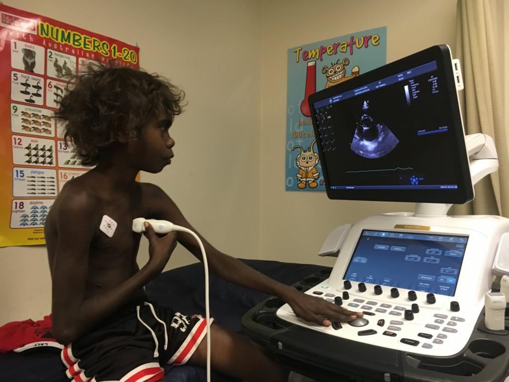
- Transactorial ultrasound is the most popular and common type of research, which proceeds by installing a sensor on the chest to visually evaluate all the indicators of the structure and work of the heart.
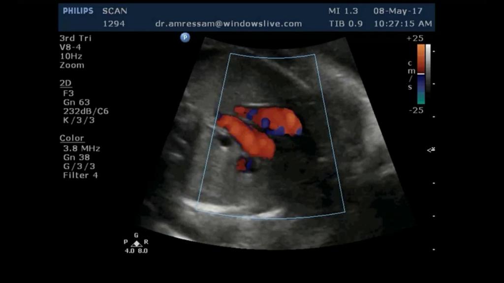
- Doppler ultrasound (echocardiography) allows you to evaluate the movement of blood in the heart and coronary vessels.
- Contrast echocardiography (ultrasound) allows the cardiologist to get a clearer picture of the inner face of the heart by introducing an X-ray contrast solution into the blood.
- The stress test combines transactor and Doppler ultrasound, is performed using physical activity or injecting drugs into the body, and allows you to identify areas on the heart where coronary artery stenosis can occur.
- Transesophageal echocardiography is performed through an ultrasound transducer, which is inserted through the throat or esophagus to enable the doctor to see the most accurate image of the heart in a moving mode.
Ultrasound examination
Now that you know where to do an ultrasound of the heart of a child or adult, and also figured out the different types of this procedure, let's determine how this diagnosis proceeds:
- On a standard ultrasound, the patient is stripped to the waist, placed on a couch on his back, turned over on his left side, after which his chest is smeared with a certain gel, and the doctor guides the sensor through it, lingering at one point or another for scanning the heart.
- The stress echocardiogram is carried out first as an ordinary ultrasound, and then the patient is given Dipyridamole and Dobutamine to cause pharmacological stress, or the person is forced to perform certain physical exercises, loading the body, and after a while they do the usual ultrasound to check how the heart copes with the load.
- Transesophageal ultrasound is performed by inserting an endoscope through the throat or esophagus into the patient's body, before which his oropharynx is irrigated with an anesthetic, which minimizes pain and discomfort.
The norm of the results of ultrasound of the heart of an adult
Now that we have figured out how Echocardiography (ultrasound) is done, let's figure out what should be the normal indicators of such a diagnosis, indicating the excellent health of an adult:
- The size of the atrium of the left should be in the range from 2.3 to 3.8 centimeters.
- The final diastolic size of the left ventricle (CRD VL) should be in the range from 3.7 to 5.6 centimeters.
- The final systolic size of the left ventricle (DAC VL) should be in the range from 2.1 to 3.6 centimeters.
- The thickness of the left ventricular wall should be between 0.8 and 1.1 centimeters.
- The thickness of the interventricular septum should be between 0.8 and 1 centimeters.
- The size of the atrium of the right should be in the range from 2.3 to 4.6 centimeters.
- In the basal section, the size of the right ventricle (right ventricle) should be in the range of 2 to 3 centimeters.
- The wall thickness of the right ventricle should be in the range from 0.2 to 0.5 centimeters.
- The size of the atrium of the left should be in the range from 2 to 3.6 centimeters.
- The transpulmonary blood flow velocity should be in the range of 0.6 to 0.9 m / s.
- There should be no fluid in the pericardial region at all, or its volume should not exceed 30 ml.
- There should not be blood clots, heart attacks and regurgitation.
The norm of the results of ultrasound of the baby's heart up to a year
But the norm of ultrasound of the heart of babies will depend on their age:
- In a baby up to 1 month, the CRC of the LC should be in the range from 1.3 to 2.3 centimeters; KSD ZhL - from 0.8 to 1.6 centimeters; wall thickness of the rear LC - from 0.2 to 0.5 centimeters; interventricular septum thickness - from 0.2 to 0.6 centimeters; the size of the atrium of the left (PL) - from 0.9 to 1.7 centimeters; the size of the ZhP - from 0.2 to 1.3 centimeters.
- In a baby up to 3 months, the CRC of the LC should be in the range from 1.6 to 2.6 centimeters; KSD ZhL - from 0.9 to 1.6 centimeters; wall thickness of the rear LC - from 0.2 to 0.5 centimeters; interventricular septum thickness - from 0.2 to 0.6 centimeters; submarine size - from 1 to 1.9 centimeters; the size of the ZhP - from 0.2 to 1.3 centimeters.
- In a baby up to 6 months, the CRC of the LC should be in the range from 1.9 to 2.9 centimeters; KSD ZhL - from 1.1 to 2 centimeters; wall thickness of the rear LC - from 0.3 to 0.6 centimeters; interventricular septum thickness - from 0.2 to 0.6 centimeters; submarine size - from 1.2 to 2.1 centimeters; the size of the ZhP is from 0.2 to 1.4 centimeters.
- In a baby up to a year, LV CRD should be in the range from 2 to 3.2 centimeters; KSD ZhL - from 1.2 to 3.2 centimeters; wall thickness of the rear LC - from 0.3 to 0.6 centimeters; interventricular septum thickness - from 0.2 to 0.6 centimeters; submarine size - from 1.4 to 2.4 centimeters; the size of the ZhP is from 0.3 to 1.4 centimeters.
Optimal echocardiography results for a child 1-10 years old
Also, according to age, the norms of ultrasound of the heart in children from one year to 10 years differ:
- In a child of 1-3 years old, the CRC of the LC should be in the range from 2.3 to 3.4 centimeters; KSD ZhL - from 1.3 to 2.2 centimeters; wall thickness of the rear LC - from 0.3 to 0.7 centimeters; interventricular septum thickness - from 0.2 to 0.6 centimeters; submarine size - from 1.4 to 2.6 centimeters; the size of the ZhP is from 0.3 to 1.4 centimeters.
- In a child of 3-6 years, the CRC of the LC should be in the range from 2.5 to 3.6 centimeters; KSD ZhL - from 1.4 to 2.5 centimeters; wall thickness of the rear LC - from 0.3 to 0.8 centimeters; the thickness of the interventricular septum is from 0.3 to 0.7 centimeters; submarine size - from 1.5 to 2.7 centimeters; the size of the ZhP - from 0.4 to 1.5 centimeters.
- In a child of 6-10 years old, the CRD of the LC should be in the range from 2.9 to 4.4 centimeters; KSD ZhL - from 1.5 to 2.9 centimeters; wall thickness of the rear LC - from 0.4 to 0.8 centimeters; interventricular septum thickness - from 0.4 to 0.8 centimeters; submarine size - from 1.6 to 3.1 centimeters; the size of the ZhP - from 0.5 to 1.6 centimeters.
Optimal ultrasound scans for heart valves
For an accurate diagnosis, it is not enough to know the norms of ultrasound of the heart, which show its size and structure, you still need to have additional information about the heart valves. Their pathology is determined with the help of stenosis and insufficiency coefficients. Stenosis is a narrowing of the valve opening, due to which the heart chamber with great difficulty passes blood through it. Insufficiency is the opposite state, indicating that the valves of the heart valve stop working, and then the blood when moving from one chamber to another returns back, which indicates a decrease in the efficiency of the heart. The coefficients of stenosis and insufficiency can vary in the region of 1-3, and the higher this figure, the more serious the pathology.
Interpretation of ultrasound findings and detection of heart disease
Correctly deciphering the results of ultrasound of the heart can only a cardiologist who collects all the data together and can, for one reason or another, determine if the patient has certain pathologies:
- With myocardial infarction, you can see a dead part of the heart.
- With pericarditis, a large amount of free fluid is observed.
- With myocarditis, an increase in the chambers of the heart and a decrease in the volume of blood ejected from the left ventricle are observed.
- With endocarditis, defects in heart valves are noticeable.
- With aneurysm, protrusion of the thin walls of the heart can be seen.
- With cardiomyopathy, an increase in the thickness of the walls of the vessels is observed.
- With heart failure, there is a decrease in the volume of blood that is ejected by the heart during organ contraction.
- With a defect in the heart valve, blood flow decreases and the size of the vascular walls changes.