The brain is a vital symmetrical organ that controls all body functions and is responsible for human behavior. Its weight in infants is not more than 300 g, with age it can reach 1.3-2 kg. A highly organized organ consists of billions of nerve cells interconnected by neural connections. The network of nerve fibers has an intricate structure and is one of the most complex formations in the human body.
Human brain anatomy
The brain is divided into two large hemispheres, the surface of which is covered with many convolutions. The cerebellum is located behind. Below is the trunk passing into the spinal cord. The trunk and spinal cord through the nervous system give commands to the muscles and glands. And in the opposite direction they receive signals from external and internal receptors.
From above, the brain is covered by a cranium, protecting it from external influences. Blood flowing through the carotid arteries supplies the brain with oxygen. If for some reason there is a disruption in the functioning of the main organ, then this leads to the fact that a person goes into a vegetative (plant) state.
Brain structure
The soft membrane of the brain consists of loose connective tissue with bundles of collagen fibers forming a complex dense network. It is closely spliced with the surface of the brain and penetrates into all crevices and grooves, includes large arterial veins that deliver oxygen to the organ.
The arachnoid membrane contains cerebrospinal fluid, which performs a cushioning function and is responsible for regulating the extracellular environment between nerve cells. A transparent thin cobweb layer fills the space between the soft and hard shell.
The hard shell of the brain is a strong thick plate consisting of paired leaves and having a fairly dense structure. It adjoins the inner smooth surface to the brain, and its upper part fuses with the skull. In the places of attachment of the plate with bones, sinuses are formed - venous sinuses without valves. The dura mater plays an important role in protecting the brain from injury.
Brain departments
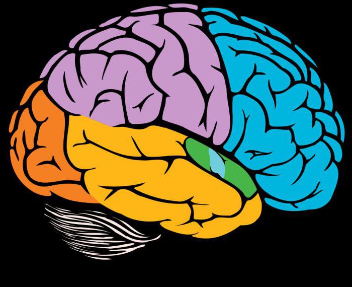
The cerebral hemispheres are divided into four zones. The picture below shows the location of the lobes of the cerebral cortex:
- The frontal part is indicated in blue.
- Violet is the parietal region.
- Red is the occipital area.
- Yellow is the temporal lobe.
Brain Department Table| Department | Where is located | Main structures | What is responsible for |
| Front (end) | Frontal lobes of the head | Corpus callosum, gray and white matter; basal nuclei - striatum (caudate nucleus, pale ball, shell), xiphoid body, fence | Behavior control, action planning, movement coordination, skills development |
| Intermediate | Above the midbrain, under the corpus callosum | Thalamus, metolamus, hypothalamus, pituitary, epithalamus | Hunger, thirst, pain, pleasure, thermoregulation, sleep, wakefulness |
| Average | Upper part of the brain stem | Quadrupole, legs of the brain | Regulation of muscle tone, the ability to walk and stand |
| Oblong | Spinal cord extension | The nuclei of the cranial nerves | Metabolism; protective reflexes: sneezing, lacrimation, vomiting, coughing; lung ventilation, respiration, digestion |
| Rear | Adjacent to the oblong section | Bridge, cerebellum | Vestibular apparatus, perception of heat and cold, coordination of movement |
The table of the departments of the brain presents the main functions of the higher organ. The slightest malfunction in the nervous system leads to serious complications and adversely affects the entire human body. Consider the most common pathologies associated with impaired brain activity.
The defeat of the basal nuclei
Basal nuclei (ganglia) are individual accumulations of gray matter in the subcortical part of the cerebral hemispheres. One of the main formations is the caudate nucleus (nucleus caudatus). A white strip separates it from the thalamus - the inner capsule. The ganglion consists of the head of the caudate nucleus, body and tail.
The main disorders with abnormal functioning of the nuclei:
- violation of coordination of movement;
- involuntary trembling of the limbs;
- the inability to learn new skills;
- inability to control behavior.
Consider the clinical manifestations of damage to the caudate nucleus.
Hyperkinesis
The disease is caused by uncontrolled spontaneous movements of a muscle group. The disease occurs against the background of damage to the nerve cells of the basal nuclei, in particular, the caudate body and the internal capsule. Provoking factors:
- cerebral palsy;
- intoxication;
- stress;
- encephalitis;
- congenital pathologies;
- head injuries;
- endocrine system diseases.
Common symptoms:
- involuntary muscle contraction;
- tachycardia;
- frequent blinking;
- squinting eyes;
- muscle spasms of the face;
- protruding tongue;
- pain in the lower abdomen.
Complications of hyperkinesis lead to limited joint mobility. The disease is incurable, but with the help of medications and physiotherapy, you can reduce symptoms and alleviate the condition of a person.
Hypokinesia
Damage to the caudate nucleus of the brain is a common cause of the development of an ailment associated with a decrease in the motor function of a person.
Symptoms and consequences:
- hypotension;
- intestinal absorption;
- impaired functioning of the senses;
- decreased lung ventilation;
- atrophy of the heart muscle;
- stagnation of blood in the capillaries;
- bradycardia;
- degenerative changes in posture.
A drop in blood pressure leads to a decrease not only in physical activity, but also in mental activity. Against the background of hypokinesia, performance is lost, and the person completely falls out of society.
Parkinson's disease
With the disease, degenerative changes in neurons occur, which leads to a loss of control over movements. Cells cease to produce dopamine, which is responsible for the transmission of impulses between the caudate nucleus and the black substance. The disease is considered incurable and is chronic.
Initial symptoms:
- handwriting change;
- slowness of movements;
- tremor of limbs;
- depression;
- muscle tension
- illegibility of speech;
- impaired gait, posture;
- a frozen expression on the face;
- forgetfulness.
If one of the symptoms appears, consult a neurologist.
Huntington Chorea
Chorea is an inherited pathology of the nervous system. The disease is manifested by mental disorders, hyperkinesis and dementia. Impairment of motor function due to gusty movements that are not amenable to human control. With the disease, a lesion of the basal ganglia, including the caudate nucleus, occurs. Although scientists have sufficient information about the anatomy of the human brain, chorea is still poorly understood.
Symptoms
- restlessness;
- sharp wave of hands;
- decreased muscle tone;
- cramps
- memory impairment;
- smacking, sighing;
- involuntary facial expressions;
- short temper;
- dancing gait.
Complications of chorea:
- inability to self-service;
- pneumonia;
- psychoses
- heart failure;
- crazy ideas;
- suicidal tendency;
- panic attacks;
- dementia.
Huntington's chorea is incurable, drug therapy is aimed at alleviating the condition and prolonging the patient's working period. To prevent complications, drugs of the antipsychotic group are used. The sooner a diagnosis is made, the less the disease will manifest itself. Therefore, at the first signs of pathology, you need to contact a specialist.
Tourette's syndrome
Tourette’s disease is a psychogenic disorder of the nervous system. The disease is characterized by motor and voice tics, which are uncontrollable.
Causes:
- damage to the structure of the brain during oxygen deficiency or during childbirth;
- maternal alcoholism during gestation;
- severe toxicosis in the first trimester of pregnancy, which negatively affects the unborn child.
Symptoms
Simple tics are short twitches of one muscle group. These include:
- mouth curvature;
- frequent blinking;
- involuntary eye movements ;
- sniffing of the nose;
- head twitching.
Complex ticks include various actions performed by several muscle groups:
- severe gesticulation;
- hyperkinesis;
- eccentric gait;
- jumping
- copying the movement of people;
- body rotation;
- sniffing around objects.
Voice Ticks:
- coughing;
- cries
- barking;
- repetition of phrases;
- grunting.
Before the attack, the patient experiences tension and itching in the body, after a seizure, this condition disappears. Drug therapy does not completely cure Tourette’s syndrome, but can reduce symptoms and reduce the frequency of ticks.
Farrah Disease
The syndrome is characterized by the accumulation of calcium in the vessels of the brain, which are responsible for providing oxygen to the inner capsule and caudate nucleus. A rare disease manifests itself in adolescence and middle age.
Provoking factors:
- carbon monoxide poisoning;
- thyroid dysfunction;
- Down syndrome;
- radiation therapy;
- microcephaly;
- tuberous sclerosis;
- violation of calcium metabolism.
Symptoms
- trembling limbs;
- cramps
- facial asymmetry;
- episindrome;
- slurred speech.
Farah syndrome is not fully understood and has no specific treatment. The progression of the disease leads to mental retardation, impaired motor function, disability and death.
Nuclear jaundice
The form of jaundice in newborns is associated with a high concentration of bilirubin in the blood and basal ganglia. With the disease, partial damage to the brain occurs.
Causes:
- prematurity;
- anemia;
- underdevelopment of body systems;
- multiple pregnancy;
- hepatitis B vaccination;
- underweight;
- oxygen starvation;
- hereditary liver diseases;
- Rhesus conflict of parents.
Symptoms
- yellowing of the skin;
- drowsiness;
- temperature rise;
- decreased muscle tone;
- lethargy;
- refusal of breastfeeding;
- rare breathing;
- enlarged liver and spleen;
- head tilting;
- cramps
- muscle tension
- vomiting
Treatment is carried out by exposure to a blue-green spectrum of rays and blood transfusion. To replenish energy resources, droppers with glucose are placed. During the illness of the child, a neurologist observes. The baby is discharged from the medical institution only with the normalization of blood counts, and the disappearance of all symptoms.
Damage to the caudate nucleus of the brain leads to severe incurable diseases. For the prevention and relief of symptoms, the patient is prescribed lifelong drug therapy.