The information in this article is intended to give a better idea of the two bones of our body, namely the sciatic and femoral. We will consider their structural features, for example, the presence of a branch in the sciatic bone or trochanter in the femur, as well as their shape and the process of ossification.
General anatomical information
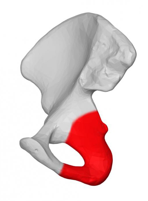
The sciatic bone is a structure in the body consisting of two elements, one of which represents its branch bent at an angle, and the second is called the body. The bone body is involved in the formation of the posterior lower acetabulum. On the back of the body there is a bone protrusion called the sciatic spine. Behind him is the sciatic notch. In the lower part, the bone body smoothly transforms into a part of the branch, which is located in the upper part of the same bone. Under the sciatic spine is a small notch of this bone, and in the opposite direction from it (on the other side) is the posterior obturator tubercle. The ischial bone of the pelvis has rough thickenings on the posterior surface of the lower part of the curved fragment of the chain, they are called the ischial tubercles. In the front of the branch fused with the lower part of the pubic bone.
The ischium has thickenings similar to those of the pubic bone. For example, a body located in the acetabulum, and branches that form an angle with respect to each other. This formation has a very thickened apex and is called the ischial tubercle.
A small sciatic notch is located along the posterior surface of the body and upward toward the tubercle. It is separated by an awn from a large tenderloin. Part of the bone moves away from the tuber and protrudes into the lower part of the pubic bone. This formation is intended to surround the obturator hole lying in the lower part medially with respect to the acetabulum. It has a triangular shape and rounded corners. A general view of the ischium in the photo is provided below.
Ossification process
The ossification of the sciatic bone occurs in four stages, which we will now consider, as well as trace the connections between them. The first period of ossification begins in a newborn baby. In his image from the x-ray machine, you can clearly distinguish 3 parts of the pelvis, which are separated by large gaps. In some places of contact between the bones of the pubis and sciatica, the lumen is not visible. This says that in these areas the bones are projected one on top of the other, and vice versa. The picture shows that they are one whole fragment, similar to claws, but not closed. After 8 years, at the second stage, the branches are combined into a single structure, and by the age of 14-16, when the third stage begins, in the area of the acetabulum the remaining branch is connected to the ilium, so they form the pelvic bone. In the interval from 12 to 19 years, points begin to form, to which muscles and ligaments will be attached. The final stage of sciatic bone ossification occurs in the period from 20 to 25 years, which is caused by their fusion with the main bone mass.
Gender differences
The structure of the pelvic bones in both sexes is different. This is due to the female reproductive function: the pelvic bones of the expectant mother must be more plastic so that the fetus passes through the birth canal. The difference in structure between the male and female pelvic bone is evident from the age of 20. Until the manifestation of sexual differences, it retains the appearance of an elongated funnel, characteristic of childhood. Sciatica synostosis in the areas of the acetabulum occurs with the help of additional bone formations. They can stay for a long time. Their x-ray clearly shows, they look like debris.
Familiarity with the structure of the femur
Based on the anatomy of the femur, it should be concluded that this is a formation represented by tubular bone tissue. Her body is in the shape of a cylinder, slightly curved in the front; along its surface there is a rough strip (linea aspera) behind, which serves as a place of attachment of muscles and tendons. In the lower part, the body begins to expand.
Anatomical description
We will begin to consider the anatomy of the femur with the proximal pineal gland. On its surface is the head of this bone (caput femoris) with the articular surface located on it, which articulates with the acetabulum. There is a dimple in the central part of the surface on the head. The connection of the head and body of the bone is clearly expressed by the neck (Cullum femoris). The axis of this formation is at an angle of one hundred and thirty degrees with respect to the longitudinal axis. The transition region of the neck into the body has two tubercles, called the greater and lesser trochanters. The first protrudes in the lateral (external lateral) direction and is easily detected through the skin. The second is located on the back of the inside. Not far from the major trochanter, a trochanteric fossa (fossa trochanterica) lies in the femoral neck. Skewers are connected in front with an intertrochanteric line, but the posterior region is combined due to a crest.
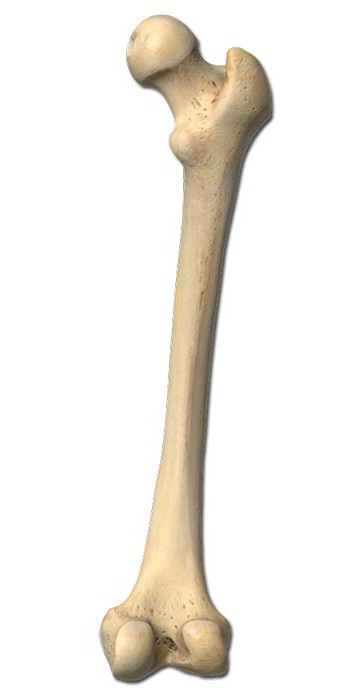
The anatomy of the femur is arranged in such a way that the distal end of its body, starting to expand, flows into the lateral and medial condyle, between which lies the intercondylar fossa (fossa intercondylaris), clearly expressed behind.
The condyles of the thigh have articular surfaces, with the help of which the joint of the femur with the tibia and patella occurs. The superficial radius of the condyles decreases from the anterior to the posterior direction, forming a spiral.
To summarize
From the above information, we can draw conclusions regarding the structure of the bones of the sciatica and thigh. Both bones belong to the bones of the lower part of our body, are very different in structural features and are formations of different types: the femur is called mixed, and the sciatic is flat. The femur, unlike the sciatic bone, has a simpler process of ossification.