Computed tomography is a special study that allows the doctor to clearly see the organ using x-ray radiation. But very often the patient is prescribed not ordinary tomography, but a contrast agent is used for CT, which allows to give the most objective assessment of the condition of internal organs, tissues or blood vessels.
Purpose of CT with contrast
As in the case of MRI with a contrast agent, computed tomography with contrast is performed by introducing certain substances into the body that improve the visibility of a particular area. So, CT of the lungs with a contrast agent allows you to better see the lungs; CT scan of the abdominal cavity makes it possible to view the intestines, stomach, pancreas, bile and liver; CT of the retroperitoneal space allows you to better examine the kidneys, adrenal glands, urinary tract, lymph nodes and blood vessels.
Such a study is carried out in cases where the doctor is important:
- visually separate nearby internal organs from the loop of the intestine;
- conduct respiratory examinations;
- visualize a tumor, cyst or inflammation of a particular organ;
- diagnose the exact condition of the blood vessels;
- determine the degree of malignancy of the tumor in the body;
- using CT with the introduction of a contrast agent to assess the condition of the internal organ before surgery;
- diagnose the course of chronic or acute pathologies in the body that cannot be detected in another way;
- monitor the patient's condition during the current treatment.
Contraindications to CT with contrast
However, this type of research is not shown to everyone. Thus, you can not enter a contrast agent for CT scan of the abdominal cavity, retroperitoneal space or lungs in cases where the risk from this study will exceed its need. Therefore, before performing computed tomography with contrast, a biochemical blood test should be done and an examination should be done so that after the doctor has analyzed all the facts, he individually prescribed a CT scan. Particular attention here will need to be paid to the patient's presence of bronchial asthma, diabetes mellitus, an allergy to seafood or iodine, and the presence of severe diseases of the kidneys, liver, thyroid gland and heart, which may be relative contraindications for the study. But the main thing - a direct contraindication to it is the presence of renal failure in the patient - in this case, the doctor can prescribe him only a CT scan without a contrast agent, because otherwise the risk of serious complications will be simply catastrophically high. In addition, the study should not be prescribed to pregnant women and young children, and nursing mothers after computed tomography should refrain from breastfeeding for a day.
Study side effects
If before a computed tomography with contrast the patient underwent a thorough medical examination, then most likely he should not be afraid of side effects, since they happen extremely rarely. However, sometimes after administration of a contrast agent for CT in a patient:
- dizziness and nausea may occur, similar to those that appear from motion sickness on a carousel;
- if the contrast was introduced by a bolus method, then at the site of puncture of the skin with a needle a slight itching and redness may occur, but this happens only in people with too sensitive skin;
- when the contrast enters the bloodstream and spreads through the blood vessels, a sensation of heat or cold may appear, which is completely normal and will pass immediately after the procedure;
- if the patient was not aware of an allergy to iodine or seafood, during the study he may experience an allergic reaction in the form of itching, redness, swelling, rashes, shortness of breath or coughing, which can be removed with antihistamines;
- in one out of a hundred people, nausea or vomiting may occur during the procedure, blood pressure may rise or loss of consciousness may occur, after which the study stops, and the doctor should begin symptomatic treatment.
Harm from computed tomography
Even if the patient is not injected with a contrast agent during CT, but just done an ordinary computed tomography, this study can cause some harm. And all because during computed tomography a person receives a considerable radiation exposure to background radiation, which is approximately 2 mSv during CT scan of the head, and about 30 mSv during computed tomography of the abdominal cavity. Such a dose of radiation is considered significant enough and can damage cells at the molecular level. And in this case, it remains to rely only on the strength of the patient’s immune system, which will either independently eliminate this damage or lead to the development of a cancerous growth. Therefore, in order not to harm yourself, it is better to consult a doctor before the study, who can accurately say about the advisability of performing a tomography.
With special care, you should check the need for computed tomography for children who are especially sensitive to x-rays due to the fact that their body develops, which means that cells divide more actively. And because of this activity, they are more susceptible to any danger, including radiation. Therefore, due to the riskiness of the procedure, CT is prescribed to children only in the most emergency cases, when there is a serious danger to their health, and other examination methods do not help.
CT scan damage
It does not matter whether the patient is prescribed a CT scan of the kidney with a contrast agent or computed tomography of blood vessels, lungs, ureters, spinal cord or any other organ, it should be remembered that the contrast does not linger for a long time inside the body, is not able to get into the tissues of organs and, therefore, completely harmless to humans. However, not all so simple. There are several situations where it is better to refrain from introducing contrast into the body, because the risk from this procedure will exceed the benefit from it.
- If the patient suffers from renal failure, then after the study he may receive toxic poisoning, since the withdrawal of the contrast agent from the body occurs through the kidneys.
- If the patient is allergic to iodine, which is the main component of the contrast, then the study should be abandoned, since an allergic reaction can occur, up to serious breathing problems.
- If the patient suffers from autoimmune thyroiditis or hyperfunction, then there is a risk of serious damage to the thyroid gland.
Classifications of contrast agents
There are different types of contrasts depending on whether a CT of blood vessels of the heart with a contrast agent, CT of the brain, peritoneal cavity, bronchi, gall bladder, or any other organs is prescribed.
- Omnipack and Urografin are water-soluble contrasts that are used to evaluate the condition of the ureters, kidneys, blood vessels, and lymph nodes.
- "Iodolipol" is a fat-soluble contrast, which is necessary in order to diagnose a disease of the bronchi, spinal cord and any spinal structures.
- Etiotrast is an alcohol-soluble contrast that is used to assess the condition of the bile ducts, gall bladder, and intracranial canals.
- Barium sulfate is a contrast that cannot be dissolved and which is used to study the digestive tract.
In addition, there are two more types of contrast agents for CT, which differ by the principle of absorption of x-rays.
- Positive are barium and iodine, which can absorb radiation much better than body tissue.
- Negative - these are gases that weakly absorb x-ray radiation, so they are used only when it is necessary to provide a transparent background with accurate detection of neoplasms. Most often, gases are introduced into the bladder.
The process of computed tomography with contrast
Now let's look at how contrast agent is introduced during CT and how this study is generally conducted. All computed tomography using contrast lasts about 30-40 minutes, of which a maximum of 5-10 minutes is allotted for the introduction of contrast, the rest of the time the doctor evaluates the data and analyzes what he sees on the screen. Contrast can be introduced into the body in three ways.
- For CT of the stomach and intestines, the patient takes the contrast agent orally, swallowing it, after which it is rapidly absorbed into the body, and because of this, the clarity of the image of the organs and tissues of the gastrointestinal tract immediately increases.
- If the clinic where the study is carried out has a first-generation device, then the contrast is injected into the vein manually, which, unfortunately, does not allow you to adjust the rate of its entry into the body.
- If the CT apparatus is equipped with a syringe, then the contrast is injected into the vein by the blues pathway, due to which the rate of entry of the substance into the body can be controlled in order to prevent side effects.
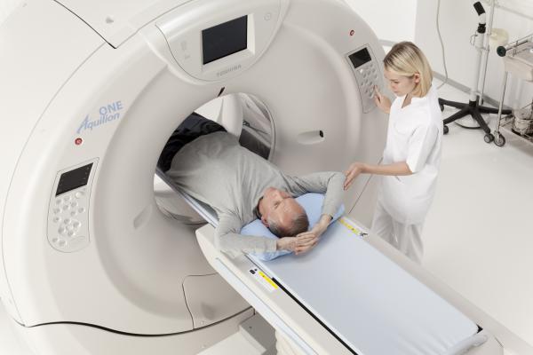
The patient himself, while scanning his body, should lie still, not move, not nervous, and sometimes hold his breath, which he learns with the help of light pointers.
PET CT with contrast agent
Separately, mention should be made of positron emission tomography, which is one of the latest modern CT methods and allows you to most accurately examine human organs, helping to detect cancer in the early stages or during its development. That is why PET CT with contrast is most often prescribed to patients preparing for the treatment of tumors of the lungs, head, larynx, tongue, intestines, liver, mammary glands and kidneys, as well as the treatment of melanoma and lymphoma. Indeed, with the help of such computed tomography, doctors manage to detect about 65% of cancerous tumors.
In addition, this type of study is prescribed for problems with memory or the nervous system, to identify foci of epilepsy, to clarify the degree of development of Alzheimer's disease, to detect the presence of the consequences of a heart attack, for coronary heart disease and to study cerebral circulation. In all these cases, tomography will help determine the treatment method and find out how effective it is.
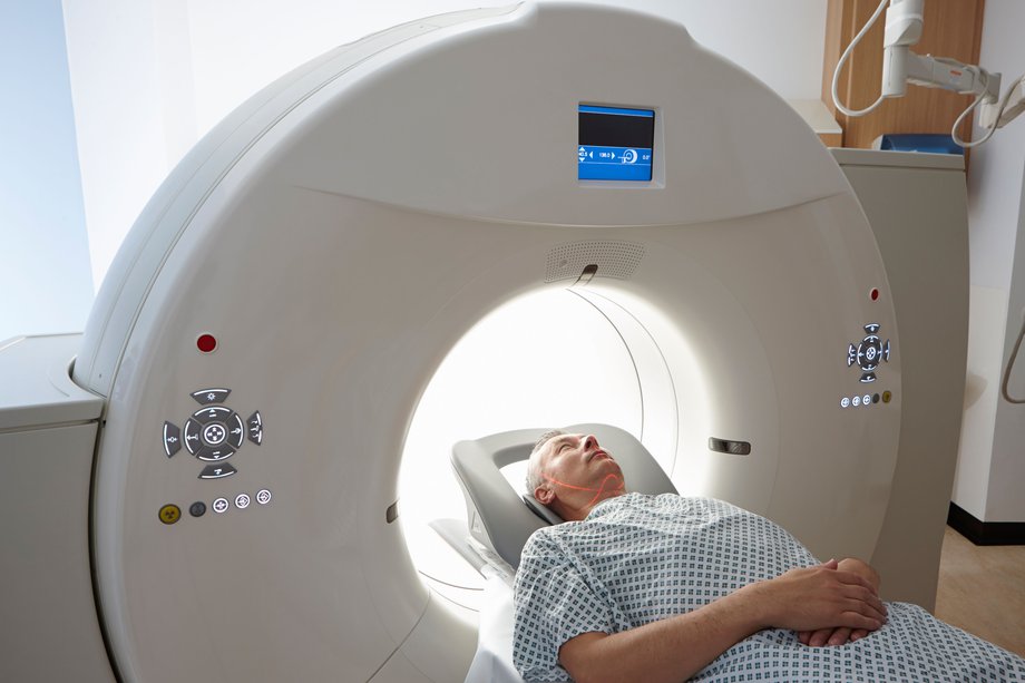
This study goes almost the same way as conventional computed tomography. True, a contrast agent for CT scan of the abdominal cavity or retroperitoneal space is injected into the vein 45 minutes before the start of the study, and all this time the patient must be silent and not move. Then the patient is placed on a moving couch and sent to the scanner, the sensors of which begin to pick up signals that will be transmitted by a tomograph to a computer screen in the form of an organ image on which the affected areas will be highlighted in color.
CT preparation with contrast medium
In order for the study to give the correct results and pass as safely as possible, you need to prepare for it. Two days before it, you will need to start a diet, abandoning products such as alcohol, fruit juices, sodas, fermented milk products and products made with yeast. And at the very moment of the study, you need to try to free your stomach from food as much as possible, so if a CT scan is scheduled for the morning, then you need to do an examination on an empty stomach, and the night before should be limited to a light dinner. If CT is scheduled for lunch, then 5 hours before the procedure, you can easily have breakfast, and if the tomography is scheduled for dinner, you can have a good breakfast, but in no case do not have lunch. And just a few hours before tomography, you will need to put yourself a cleansing enema or take a mild laxative to empty the intestines.
And after the examination, it was recommended to eat more apples, seaweed, almonds, lentils, pumpkins, oats, walnuts and beans to get rid of the received dose of radiation.
Computed Tomography Results with Contrast
And now, when we know how the contrast agent is introduced during CT scan of the abdominal or retroperitoneal space, what are the contrasts and what are the indications or contraindications for such a study, let's find out what we can find out after computed tomography. So, after a CT scan, the doctor will be able to detect in the patient:
- benign or malignant tumors, as well as determine how much they have grown into nearby tissues;
- chronic or acute liver damage;
- stones in the ureters or in the kidneys;
- CT of vessels with a contrast medium allows to detect various vascular pathologies, including atherosclerosis;
- foreign bodies and cystic formations;
- problems with the outflow of bile and the presence of stones in the bile ducts or gall bladder;
- inflammation of the internal organs.