Along with the development of the pharmacological branch of medicine and the discovery of new data on the structure of the body, a huge role is played by the improvement of innovative technologies that greatly facilitate the work of a doctor. So, these are various methods of radiation (fluorography, x-ray, computed, magnetic resonance or nuclear magnetic tomography, angiography), as well as direct visual diagnostics, which includes various soundings (fibrogastroduodenoscopy, esophagogastroduodenoscopy, uterine probe, coproscopy, capsule video endoscopy, duodenoscopy sounding) , etc.
Method Features
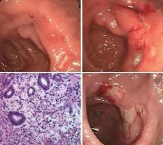
Visual methods allow the diagnostician directly in real time to see the condition of many internal organs of a person. First of all, this applies to most sections of the food tube, that is, the pharynx, esophagus, gastric cavity, small and large intestines, as well as the bile ducts and the gall bladder itself are subject to inspection. Thus, it is possible to directly examine the state of these organs, identify erosion, inflammatory or ulcerative necrotic changes, tumor processes, as well as evaluate the degree of treatment effectiveness using the obtained morphological picture or guide them on the operation. However, the abilities of these methods are not limited to this, because with the help of probes you can also deliver medicine precisely, take portions of gastric or bile juice for analysis, as well as biopsy samples from various departments. Thus, the doctor will be able to get a complete picture of what is happening in the body, timely make the correct diagnosis and correctly prescribe treatment.
The benefits of endoscopy
Esophagogastroduodenoscopy (EGD) is one of the methods of sounding that allows you to examine the esophagus, stomach and duodenum with a micro camera. Of course, there is another method, non-invasive - X-ray examination after taking barium suspension in the form of porridge for contrasting the mucous membrane. However, this method has rather limited capabilities and allows you to see in the picture except chemical burns, tumor processes, ulcers and erosion. While the examination of endoscopy can reveal bleeding, take biopsy samples for microscopic analysis and examine the organs in more detail. Also, this technology allows you to assess the condition of the departments after the
surgery, namely healing, the integrity of the joints, bruising, remove foreign objects, diagnose varicose veins of organs.
The essence of endoscopy
Many patients who received a referral from their attending physician for this examination often ask the question: "endoscopy, what is it?" The abbreviation usually scares patients and even leads to a dead end for those who have never before encountered visual diagnostics. So let's try to figure out how it works. The endoscopy of the stomach or other gastrointestinal tract is carried out using a special probe - a flexible controlled tube, consisting of an elastic material and having additional fiber optics (a kind of camera and light bulb) and manual control technique. This allows not only to lower it along the gastrointestinal tract, but also to turn it to the sides or down to get a complete visual picture. After the examination is completed, the probe is carefully removed so as not to create iatrogenic mechanical damage to the internal membranes of the organs.
Precautions during the procedure
Since most patients, especially children, are still afraid of this study, the doctor must explain to each of them before endoscopy that it is an unpleasant, but very necessary method to maintain their health. He should be told that there are probes of different diameters, it takes a little time (literally 20-30 minutes), and immediately before it is injected with novocaine in the form of an aerosol, the oral cavity is anesthetized. It is also necessary to suppress the
vomiting reflex, since the probe will irritate the
palatine tongue and pharyngeal baroreceptors. Therefore, if the patient feels a bitter taste in the mouth or a slight swelling of the tongue, this is an absolutely normal reaction to the drug. In addition, after anesthesia, a mouthpiece is inserted - a sterile plastic device for protecting the lips and teeth of the patient during the introduction of the probe into the digestive tract.
Additional research
In parallel with conducting a direct examination, patients with diseases of the cardiovascular system are cuffed to control blood pressure and an ECG is monitored, and in patients with respiratory failure in the diagnosis, a pulse measurement is also performed. Before radiation methods of research, there are other advantages in endoscopy. Preparation for the latter is practically unnecessary and only requires an empty stomach if it is early in the morning, or 6-24 hours after the last meal, the time depends on the preliminary diagnosis. So, anxious patients with hypersecretion of hydrochloric acid in the stomach require a longer preparation period, from 12 to 24 hours, as well as additional intravenous administration of sedative and analgesic drugs.
Warnings and recommendations
At the same time, you should ask the patient in advance to come with an escort, and also warn him about the endoscopy method that this can become quite traumatic for his internal organs if he does not follow the doctor's recommendations on the technique of the procedure. And also immediately before the examination, it is necessary to remove contact lenses, dentures, tight clothing. In addition, the patient should be reminded that he will have excessive salivation during endoscopy, that this is also a normal manifestation of the body's reaction, and therefore should not be prevented. And to collect saliva, they put a clean towel (often brought from home) under the cheek of the subject, and if necessary, apply an electric suction pump.