Myocardial infarction is a dangerous heart disease, which if failure to provide timely assistance leads to the death of the patient. Its essence lies in the fact that as a result of problems with the vessels, namely, their blockage by blood clots, blood flow to the heart tissues is disrupted, and their necrosis, that is, death, occurs. Competent medical care provided in the first half hour after the onset of signs of a heart attack can save the patient's life. An important function in assisting is assigned to the correct diagnosis, which is impossible without laboratory diagnosis of myocardial infarction.
Research methods for myocardial infarction
If myocardial infarction is suspected, the first thing to do to save the patient is to call an ambulance. Indeed, 30-40 minutes after the cessation of blood flow to tissues, their death begins, which is not amenable to treatment and can be fatal.
After the patient enters the medical institution and provides him with first aid, doctors resort to a series of manipulations aimed at confirming or refuting the diagnosis. We can distinguish laboratory and instrumental diagnosis of myocardial infarction.
Diagnosis methods used:
- general blood analysis;
- blood chemistry;
- troponin test;
- Analysis of urine;
- electrocardiography;
- radiography;
- Ultrasound of the heart (echocardiogram);
- myocardial scintigraphy;
- coronarography;
- Magnetic resonance imaging;
- history taking;
- palpation;
- percussion;
- auscultation;
- body temperature measurement;
- blood pressure control.
Among all the methods, laboratory diagnosis of acute myocardial infarction plays a leading role, since in the presence of damage to heart tissue, the composition of the blood changes significantly.
Blood biochemistry
With myocardial infarction, death of the muscle cells of the heart occurs, which leads to the ingress of special substances into the blood, the concentration of which in the blood of a healthy person is minimal. To diagnose myocardial infarction, it is necessary to determine the content of these specific markers.
Substances indicating cardiac muscle necrosis:
- CPK (creatine phosphokinase) is a nonspecific marker characteristic of any minimal muscle damage;
- MV-KFK (creatine phosphokinase-MV) - can also be increased with other injuries, so it is worth considering that the presence of this substance will indicate a myocardial infarction with a sharp increase in its concentration;
- myoglobin - a muscle enzyme, the amount of which in the blood exceeds the norm by 10 times within 4-8 hours after the start of the death process;
- aspartate aminotransferases - appear no earlier than 8 hours after necrosis, are more likely to help in the diagnosis of large focal heart attack;
- alanine aminotransferases - the amount increases one day after the attack, an increase in concentration is also characteristic of liver problems;
- lactate dehydrogenase - the maximum amount of a substance is observed only 6 days after myocardial infarction;
- troponin-T is a cardiospecific marker, which is determined in the blood already 2-3 hours after the attack, its amount is constantly increasing and reaches its maximum mark 10 hours after the heart attack, while up to a week it can remain at a fairly high level;
- troponin-I is a protein that appears in the blood almost immediately after the onset of necrosis, which allows you to diagnose myocardial infarction at an early stage.
Due to its specificity, troponins can not only diagnose an early heart attack, but also predict the likelihood of a patient surviving, choose the optimal treatment regimen, and with a timely study and predict the occurrence of an attack.
General blood analysis
In addition to analysis for biochemistry, laboratory diagnosis of acute myocardial infarction includes a general blood test. Although this method does not reveal specific markers, but thanks to it, it is possible to identify signs characteristic of the inflammatory process.
OAC indicators in laboratory diagnosis of myocardial infarction:
- an increase in ESR (erythrocyte sedimentation rate);
- leukocytosis (high white blood cell count);
- change in blood count (stab shift);
- aneosinophilia (lack of eosinophils);
- an increase in platelet count if the attack is triggered by a blood clot and increased blood clotting;
- an increase in the number of white blood cells due to an increase in the content of neutrophils.
To increase the likelihood of a favorable prognosis, it is important to conduct all studies in the first hours after the first symptoms of the disease.
Express method
To facilitate laboratory diagnosis of myocardial infarction, especially with the uncertain results of other studies, the troponin test is widely used, which is an express method in determining cardiac muscle necrosis.
It is a test strip with a special indicator, on which it is necessary to apply a few drops of the patient's blood, after which it remains to evaluate the result in the next window of the test. Two colored stripes indicate confirmation of the diagnosis, one refutes the assumption of necrosis.
Analysis of urine
Urinalysis is not a specific research method in the diagnosis of myocardial infarction and angina pectoris. This technique is used to detect concomitant complications.
The following indicators indicate complications in the work of the kidneys:
- leukocytosis;
- traces of blood;
- the presence of red blood cells.
With a decrease in the amount of urine released per day, we can talk about the accumulation of decay products of heart muscle tissue.
The Importance of an Electrocardiogram
Diagnostic methods for myocardial infarction are not limited to laboratory tests. Moreover, without examination and instrumental diagnosis, it would be very difficult to make a diagnosis.
One of the methods for making this diagnosis is electrocardiography - a technique that allows you to quickly and accurately obtain data on the work of the heart muscle. In the event of necrosis using an ECG, you can:
- confirm the diagnosis;
- determine the stage of the lesion;
- identify the place of occurrence of necrosis;
- choose the best treatment methods;
- predict complications.
Stage of myocardial infarction:
- Sharp. The first stage of the pathology, which is diagnosed 1-3 days after the attack.
- Subacute. It can continue up to 7 weeks after the onset of necrosis, but, as a rule, ends after 3 weeks.
- Scarring. The third stage does not have clear boundaries in time; on average, it lasts up to 3 months.
Most accurately it is possible to diagnose large focal lesions, that is, tissue death, significant in area. In other cases, as well as with various features of the course of the disease (for example, if chronic scars are present), problems may arise in the diagnosis of myocardial infarction, clinical recommendations in such situations require other research methods.
Radiography
An X-ray of the chest organs is used not so much for the diagnosis of myocardial infarction, but for the determination and prediction of complications after an attack.
R-graph shows:
- the presence or absence of pulmonary edema, which is characteristic of heart problems;
- blood flow in the lungs and pulmonary artery;
- clarity of vascular pattern;
- stratification of the walls of the aorta.
The accuracy of x-ray with necrosis of the heart tissue does not exceed 40%, therefore, with myocardial infarction, the clinic and diagnosis of which often causes a lot of controversy, relying only on this method is unacceptable.
Patient examination
In addition to laboratory methods for diagnosing myocardial infarction and the above instrumental, other methods of making this diagnosis are widely used in medicine.
Examination of the patient is the simplest method of research, which the doctor can begin immediately after arriving at the call. Without requiring additional devices, this technique includes a number of manipulations aimed at recognizing necrosis of the heart muscle in the early stages.
Physical examination methods as part of the clinical diagnosis of myocardial infarction:
- History taking. To get a complete picture of the disease, the doctor needs to collect an anamnesis - to get information about the symptoms of the current state, past diseases in the past.
- Palpation method. To check the correct operation of the heart muscle, palpation of the myocardial point is carried out, the displacement of which indicates violations in the functioning of the heart. Using the same method, the patient’s pulse rate is measured, the lymph nodes are checked for hypertrophy.
- Percussion or tapping of the chest to determine the heart borders. The high diagnostic accuracy of the method lies in the fact that during myocardial infarction, dilatation of the left ventricle of the heart is observed (it increases in volume), which causes a shift in the borders of the heart to the left side.
- Auscultation. This procedure involves listening to the patient’s chest with a stethophonendoscope. In this case, the rhythm of the heart, the presence of noise, weakened tones, which is characteristic of a heart attack, are assessed.
- Body temperature measurement. As a rule, with myocardial infarction, the patient has a low-grade fever in the range of 37.1-37.4 ° C.
- Changes in blood pressure can also indicate myocardial necrosis. Since pumping function of the heart weakens due to necrosis, pressure drops by an average of 10-15 mm Hg. Art.
Laboratory methods for studying myocardial infarction can show a complete picture of the disease, but most often they cannot be carried out immediately after an attack. In situations where the diagnosis must be made as soon as possible, a patient examination comes to the rescue.
Ultrasound of the heart
Ultrasound examination of the heart or echocardiogram (ECHOCG) is a mandatory diagnostic method for myocardial infarction and any other heart diseases. Its importance is due to the high accuracy of the research.
Using the EchoCG, you can set:
- the degree of damage to the heart muscle and heart bag (pericardium);
- location of necrosis;
- malfunctioning heart valves;
- the state of the vessels supporting the work of the heart muscle;
- the presence of blood clots in them;
- pressure in the chambers of the heart.
Modern ultrasound machines allow not only to see the state of the heart at the current moment, but also to assess the likelihood of complications and make a forecast of the patient's quality of life in the future.
Other research methods
Laboratory diagnosis of myocardial infarction, supplemented by instrumental methods, almost always gives a complete picture of the course of the disease and its complications. But sometimes other methods are used to clarify the diagnosis:
- Myocardial scintigraphy. The technique consists in visualizing the location of necrosis of heart tissue. For the procedure, radioactive isotopes are introduced into the bloodstream of the patient, which tend to accumulate in places of tissue necrosis. The procedure is carried out in cases of difficulties in decoding the results of the electrocardiogram.
- Coronarography is a radiographic examination of a person’s blood flow, in which a contrast agent is introduced into the vascular system. The procedure can entail a number of complications (bleeding, allergic reactions, infection, decreased vascular patency). In this regard, patients who are assigned this study are approached very selectively. Indications for coronary angiography can be serious heart defects, acute heart failure, cardiogenic shock, or frequent angina pectoris after MI.
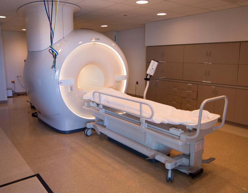
- MRI Magnetic resonance imaging is of high diagnostic accuracy and allows you to recognize the smallest areas of damage to the tissues of the heart muscle, as well as to see blood clots and generally assess the condition of the vessels. Unfortunately, this procedure is rarely used in modern medicine because of its high cost.

Diagnosis at different stages of the disease
The success of the diagnosis and treatment of myocardial infarction directly depends on the correct determination of the stage of the disease. At each of them, research indicators will differ dramatically.
Signs of myocardial infarction characteristic of the acute period:
- severe pain and burning sensation behind the chest;
- pain is also felt in the left hand;
- arrhythmia (heart rhythm disturbance);
- dizziness;
- ECG shows obstruction of the coronary vessels;
- Laboratory diagnosis of myocardial infarction records an increase in the amount of myoglobin by 15-20 times.
During the acute stage:
- the formation of a Q wave is noted on the results of the electrocardiogram, the amplitude of the ST segment changes;
- erythrocyte sedimentation rate and the number of leukocytes in the blood increase, the leukocyte formula shifts to the left;
- the number of eosinophils is growing;
- blood biochemistry is characterized by an increase in troponins T and I, as well as proteins KFK and KFK-MV.
Characteristic signs of a subacute stage are:
- the appearance of clear contours in the area of ischemia;
- decrease in myoglobin and creatine phosphokinase according to the results of a biochemical blood test;
- high troponin T and troponin I.
The last stage is characterized by the formation of a scar at the site of the lesion. Recovery from a heart attack can last up to 6 months, after which the performance of the organ will remain reduced due to a decrease in the number of cells capable of performing its function. To prevent myocardial infarction or its relapse, it is necessary to prevent the ailments of the cardiovascular system.