Eyelids (in Latin "palpebra") - a movable muscle fold in vertebrates and humans, which covers and protects the eyes. The histological structure of the layers of the structure of the eyelids is the skin, eye muscle of the eye, gland, conjunctiva.
The structure that supports the normal skeleton of the eyelid is plaque, that is, a plate about 1 mm thick. It consists of fibrous connective tissue. In its inner part there are sebaceous and sweat glands, the secret of which goes to the edge of the eyelid. By the way, the structure of the upper eyelid is such that it is larger than the lower in size.
On the inside, the eyelid is hidden by the conjunctiva. From the edge there is short, stiff hair, located in 2 or 3 rows, and maybe more - eyelashes, which include the mouth of the glands mentioned.
Hibernation present on the medial side (the so-called lacrimal papilla) is the exit site from the lacrimal canal (lacrimal apparatus).
Function
The structure of the eyelids is created in such a way as to protect the eyeball from injuries and to regulate the flow of light into the eye. Their function also lies in the distribution of tear fluid on the surface of the eyeball.
Damage to the conjunctiva causes the conjunctival reflex that closes the eyelid (the reflex arc, the medial part is the optic nerve, and the centripetal is the facial nerve).
When we talk about eye diseases, we usually think about the pathology associated with the eyeball, optic nerve or intracranial structures. The protective structure of the eyelids seems so natural that we do not pay any attention to it as long as it functions correctly. Meanwhile, his role in the process of our vision is not insignificant. It would seem that the eyelids give the impression of pieces of moving skin, but in order to have a protective function for the eyeball, this is too little. Hence their design is not too simple and functions that we still are not fully aware of.
Why do we need eyelids?
The fact that they can in some way cover or open the front of the eyeball, protecting it from drying out and injuries, is obvious to everyone. Just in case, in addition to the intentional movements of the eyelids and deliberate blinking, we also have a physiological instinct of blinking. Of course, we can blink reflectively, for example, from surprise or horror, but the main task of this act of rapid movement of the eyelids in a vertical plane is to ensure a constant distribution of the tear film on the surface of the eyeball. Even such a prosaic part of our body as the eyelids can sometimes be important (physiologically, both centuries must be the same on both sides). Its size is affected by:
- muscle tension, lifting the eyelid (levator of the upper eyelid and thyroid muscle);
- eye muscle of the eye that closes the eyelid;
- size of the eyeball and its position in the orbit.
The main muscle that causes the rise of the upper eyelid is the levator, whose function is always associated with the contraction of the upper rectus muscle (hence the movement of the eyelids up).
Eyeball Protective Functions
Eyelids are found only in vertebrates, and in humans. Already in the womb during the first 4 weeks of life of the fetus, the eyes are covered only with a thin layer of superficial ectoderm cells. However, already at the 5th week, a fold forms around the eye, at the beginning of the circular, and then with the marking of the horizontal line of the upper eyelid and its lower part, located above the center of the cornea. These two parts of the fold will become upper and lower eyelids in the near future due to the intensive differentiation of their internal structures. The four-month-old fetus already has eyelashes with sweat glands (Mall), sebaceous (Zeiss) and thyroid (Maybom). Finally, shaped eyelids include subcutaneous tissue, eyelid muscles and plaques along with the orbital septum.
Structure
The skin of the eyelids is very thin, and the subcutaneous tissue is loose and sparse, so it is easy to get swelling here. The eyelids have a grayish, thin sulcus separating two eyelids: the outer one, consisting of skin and striated muscles (in the upper eyelid and lower), and the inner, consisting of the eyelid and conjunctiva. Of particular note is the construction of compacted fibrous fabrics and elastic fibers. They give shape to the eyelids attached by a ligament to the bone edge of the orbit and, thus, together creating an orbital septum that closes the orbital entrance in front. It is there that the sebaceous glands of meibomas are located, the secret of which is to lubricate the edges of the eyelids, to close them.
Where do tears come from?
The location of the lacrimal system, whose task is to moisturize the cornea and conjunctiva, only partially affects the eyelids. But it is impossible not to mention this. Above the angle of the lateral eyelid is the lacrimal gland with tubes that divert its product into the superior conjunctival arch. There are also very small additional lacrimal glands in this area. In turn, the tear drainage paths begin near the medial angle, where on the posterior edge of each eyelid there are lace-edge spots of tears, leading them first to the wall, and then through the nasolabial tract to the lower nasal cavity. On this rather long and at the same time inaccessible road, a number of pathologies can form, ranging from inflammation, of varying severity to disturbances from injuries.
Why are we blinking?
Blinking is one of the most well-known reflex actions, and perhaps it can also be intentional. Reflexive blinking is a protective action, when, for example, a provocation is too bright light or it can be a reflex of the cornea as a reaction to the danger to the eyes. It should be noted that even all animals can blink, except for those with eyes without eyelids (for example, fish or snakes). Some animals can blink only with one eye, for example, hamsters. Sharks do not blink at all, but instead look away. The structure of the eyelid of a person’s eye allows people to blink on average 17 times per minute, and children under the age of 15 do this more often. According to statistics, we blink 14,280 times a day and 5,200,000 times a year. We blink more often when we are in tension, and less often when we absorb information.
Eyelid infections
Infections can occur repeatedly, depending on the type of pathogen, the location of the lesion, and local factors. These are minor injuries, seborrhea, excessive activity of sweat glands.
Types of disease
Purulent skin diseases of the structure of the eyelids are most often caused by Staphylococcus aureus (staphylococcus aureus) and pyrogenic streptococcus (streptococcus pyogenes). These bacteria affect the hair follicles of the eyebrows, less - the sebaceous glands. Even minor injuries in the eyelids can cause eyelid disease. The initial site of the onset of the disease can also be inflammation of the paranasal sinuses, inflammation of the internal tissues of barley or orbit. The penetration of the inflammatory process deep into, for example, along the capsule of the hair causes pain, swelling of the skin and subcutaneous tissue of the eyelids. If a causal therapeutic treatment is not performed on time, the deeper layers may become inflamed. Dangerous complications of such local infections can even include thrombosis of the cavernous sinus or purulent meningitis.
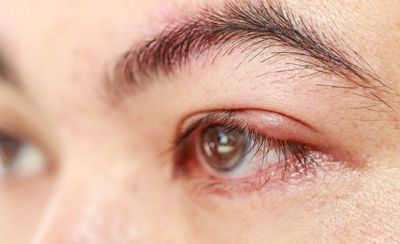
The structure of the human eyelid is so vulnerable that organs often infect purulent diseases of the glands. For example, internal barley (obstructive and purulent thyroiditis meibomas) and external barley (purulent inflammation of the Zeiss sebaceous gland and hair follicle). Inflammation here takes the form of a painful thickening located on the edge of the eyelid, sometimes bursting with the release of a purulent secretion. For treatment, warm compresses, massage, which helps to empty the lesion, eyelid hygiene, less often antibiotic treatment are used. Barley usually disappears after a week, spontaneously.
However, the disease often develops into chalazion, which is a chronic granulomatous inflammation. With it, pus accumulates in the sebaceous glands, enclosed in a clogged drainage line of the thyroid gland, under the influence of pressure penetrates into the surrounding tissues, causing a chronic sterile inflammatory reaction there. An outlet node is a nodule usually located around the drainage tube of the meibom gland near the back of the eyelids. Inflammation is slowly self-limiting, the granuloma gradually dissolves, because the contents merge through the skin or through the scalp. The process occurs with a certain improvement, if you use warm compresses, massage and antiseptic agents. In the absence of signs of resorption, chalazion should be removed surgically. Recurrent lesions of this type are indications for a histopathological examination to exclude meiboma gland cancer.
Conclusion
The proper functioning of many different parts of the eye, including the eyelid, is necessary for proper vision. However, when this is not so, we have advanced technologies that can correct the features of the structure of the eyelids. The next time you wake up in the morning, think for a moment about the miracle of your eyes that show you the world every day.