Doctors recommend undergoing a fluorographic examination once a year. This diagnostic procedure is included in the compulsory medical examination program. It helps to identify the initial signs of dangerous pathologies such as tuberculosis and cancer. Does fluorography show smoking? This question is especially often asked by young smokers. They fear that according to the results of the examination, parents will guess about their bad habit. What does the x-ray image show? And is it possible to determine from it that the patient smokes? Let's try to figure it out.
The essence of the method
Does fluorography show cigarette smoking? To answer this question, you need to understand the essence of this diagnostic method.
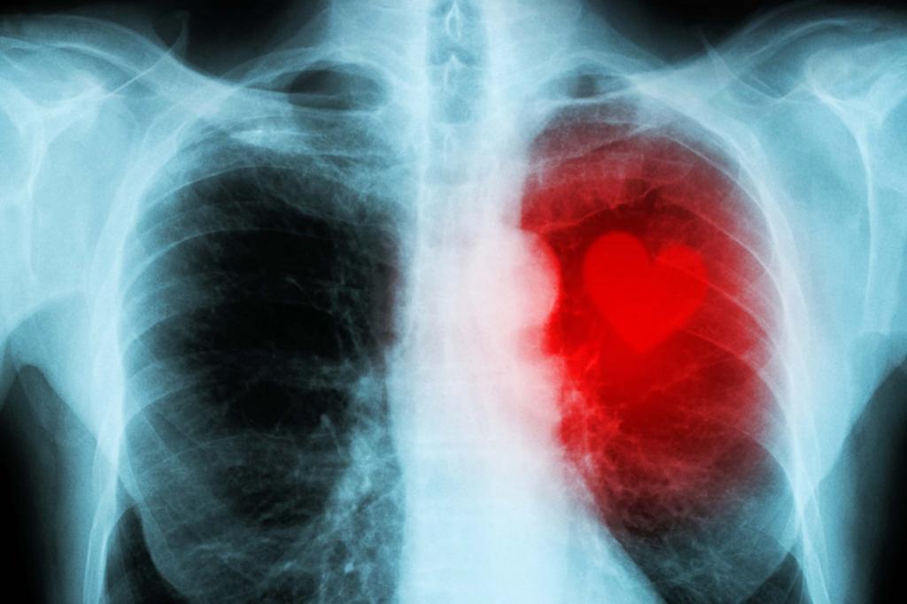
Fluorography is a type of X-ray examination. Today, diagnostics are carried out using special digital devices, which allows the use of lower radiation doses. However, the essence of the method remained the same. X-rays are passed through the patient's body, which are unevenly absorbed by the lung tissue. The image of the bronchi and lungs is displayed on a fluorescent screen and a picture is taken.
Classical x-rays of the lungs and fluorography differ in the doses of x-rays needed for the study. In conventional radiography, the patient is more exposed to radiation. For this reason, the resolution of the image is higher than with conventional fluorography.
Fluorography shows only gross and pronounced changes in the respiratory system. This is a safer but less reliable method. It is used during preventive examinations. As for the classic X-ray, this examination is often used to diagnose diseases. It shows a clear and accurate picture of pathological changes.
What the snapshot shows
During fluorography, the condition of not only the lungs, but also other organs of the chest (bones, heart, blood vessels) is examined. In the picture you can see the following features of the structure of tissues:
- structural changes;
- accumulation of gases and liquids;
- seals in the organs.
Does fluorography show smoking? This study cannot establish the very fact of a bad habit in a person. From the picture it is impossible to determine whether the patient smokes or not. But, as you know, nicotine and tobacco tar have an extremely negative effect on the respiratory system. If the patient has already had pathologies of the lungs and bronchi on the background of smoking, then a fluorographic image will show these changes.
Is it possible to identify a smoker
If a person actively and regularly smokes, then sooner or later it affects the state of the respiratory system. Radiological signs of the disease appear, which doctors call "chronic smoker's bronchitis."
However, the cause of such bronchitis can be not only nicotine. The picture can determine the presence of pathological changes, but it is impossible to establish their exact etiology. To suggest a possible cause of bronchitis, the doctor needs to collect an anamnesis. To establish the fact of smoking is possible only during a conversation with the patient.
Beginning tobacco lovers often ask the question: "Does fluorography show smoking if you smoke a year?" At such an early stage, chronic bronchitis does not occur in all patients. However, much depends on the initial state of health and the number of cigarettes smoked. In some cases, heavy smoking can lead to bronchitis within 6-12 months.
Hookah and electronic cigarettes
Many people mistakenly believe that smoking a hookah is practically harmless. However, hookah tobacco also contains toxic substances that irritate the respiratory system. Of course, the harm from regular cigarettes is much greater. However, it is impossible to talk about the complete safety of the hookah.
Does fluorography show smoking hookah? Research will only reveal the consequences of this habit. Chronic tobacco smokers also develop chronic bronchitis over time. In addition, intense tightening leads to a decrease in lung volume.
Nowadays, a lot of people use electronic cigarettes. You can even hear the opinion that with their help it is much easier to quit smoking. But is it harmless? The composition of the liquid for refueling electronic cigarettes includes various flavors. These substances can provoke the development of obliterating bronchiolitis. This is inflammation and narrowing of the small bronchi (bronchioles).
Does fluorography show smoking electronic cigarettes? If the patient has already developed bronchiolitis, then the obstruction of the small bronchi will be noticeable in the picture. In advanced cases, the examination will show the presence of cicatricial changes in the tissues. Over time, such a pathology can lead to a decrease in respiratory function and oxygen deficiency in the body.
Signs of the effects of smoking
As already mentioned, with prolonged and active smoking, changes in the respiratory system occur. Let us consider in more detail the difference in the fluorographic image of a smoker with experience and a healthy person:
- The presence of seals. Normally, the image should not have foci of dark color in the lungs. When smoking, tissue elasticity in certain areas decreases. These seal areas look like blackouts.
- Volume of the heart. In a healthy person, the size of this organ remains within normal limits. When smoking, respiratory function is severely impaired. This leads to the expansion of the heart. The organ looks a little enlarged in the picture.
- Vascular pattern. Smokers in the picture have a more pronounced network of blood vessels than healthy people. This is due to the fact that exposure to nicotine and combustion products leads to the formation of growths on arteries and veins.
- Spots on the lungs. Tobacco resins clog pores in the lungs. Areas with reduced gas exchange are formed, which appear in the image as dark or light inclusions. Normally, spots should not be detected in the image.
- Pulmonary pattern. In smokers, the shadows from the vessels in the picture are not clearly expressed. In this case, doctors say a weakening of the pulmonary pattern. This symptom indicates the onset of fibrotic changes in the tissues.
- Thickening of the walls of the bronchi. This is a result of persistent irritation of the airways with resins and nicotine. This feature indicates the presence of chronic bronchitis.
However, even by such signs, it is impossible to draw an unambiguous conclusion that the patient smokes. After all, such changes are found in people without bad habits. To establish the exact etiology of the abnormalities, doctors prescribe additional examinations.
Smoking before the procedure
Does fluorography show smoking if you smoke just before the procedure? This question is often of interest to patients. By itself, a smoked cigarette will not affect the results of the study. In the picture, only those changes in the lungs and bronchi that are already in the smoker will be visible.
Many diagnostic procedures include quitting smoking some time before the examination. However, in preparation for fluorography, this rule is not mandatory.
Conclusion
It is impossible to give an unambiguous answer to the question of whether fluorography indicates smoking. This examination cannot determine the fact of addiction to tobacco. But it accurately diagnoses the negative effects of nicotine use. Therefore, if a smoker shows pathological changes in the picture, then, most likely, they are provoked by a bad habit. This means that you need to reconsider your lifestyle and stop smoking immediately.