The upper respiratory tract is a part of a multicomponent respiratory system that absorbs oxygen from the environment, transfers it to tissues, oxidative reactions in tissues, transfers carbon dioxide to the lungs and removes it to the external environment.
Upper Respiratory Function
Anatomically, the respiratory apparatus consists of the airways (respiratory) tracts and the respiratory section of the lungs. The respiratory tract performs primarily an air-conducting function, gas exchange occurs in the respiratory lungs - venous blood is enriched with oxygen, and excess carbon dioxide is released into the alveolar air.
The respiratory tract is divided into upper and lower sections. The upper respiratory tract is the nasal cavity, nasopharynx, and oral part of the pharynx. The lower respiratory tract is the larynx, trachea, extra- and intrapulmonary bronchi.
The mucous membrane of the respiratory tract performs a barrier and protective function in the same way as all integumentary epitheliums of organs in contact with the external environment. The upper respiratory tract is a kind of calorifer-cleansing communication. Here, the inhaled air is heated, cleaned - toxic substances and foreign particles are removed from it, and moistened. Inhaled air is effectively cleaned due to the fact that the airways are lined with ciliated epithelium, and mucus secreted in the walls of the gland.
So, the airways perform the following functions:
- air delivery to the respiratory department of the lungs;
- cleaning, warming, humidifying the air;
- barrier protective;
- secretory - secretion of mucus.
The physiology of the respiratory system (as a science) studies the transport of respiratory gases under different conditions and the nervous mechanisms of breathing regulation.
The structure of the mucous membrane and the role of mucus in the respiratory tract
The mucous membrane of the upper respiratory tract has a multirow ciliated epithelium, which contains cells that are different in function and shape:
- ciliated - have ciliated cilia;
- goblet (secretory) - secrete mucus;
- microvilli (in nasal passages) - chemoreceptor (provide sense of smell);
Basal cells - cambial cells, divide and turn into goblet or ciliary.
Mucus is produced in secretory cells called goblet cells. Cells accumulate mucinogen, a substance that actively adsorbs water. Due to the accumulation of water, the cells swell, mucinogen turns into mucin - the main component of mucus. Swollen cells look like a glass - in the narrow part remains the nucleus, in the expanded part - formed mucus. When too much mucus accumulates, the cell walls are destroyed, the mucus enters the lumen of the external nose and pharynx, manifesting as mucous discharge from the nose. Mucus is also secreted in the lower parts of the respiratory system, which is manifested by a productive - wet cough.
Mucus covers the epithelium of the respiratory tract with a layer of up to 7 microns. Up to 0.75 ml of this secret per 1 kg of body weight is secreted per day in a healthy person, that is, if a person weighs about 60 kg, the volume of nasal secretion will be approximately 45 ml. During inflammation of the nasal mucosa, the volume can increase to one to two liters.
Mucus contains factors of non-specific and specific protection, due to which it has antiviral and antibacterial effects. In addition, a layer of mucus protects the lining of the respiratory tract from various injuries: thermal, mechanical, due to a change in the chemical composition of the air or its humidity.
Air purification mechanism
The upper respiratory tract is a system that effectively cleanses the inhaled air. Air purification is especially effective for nasal breathing. During the passage of air through rather narrow nasal passages, vortex movements occur. Large particles of air dust hit the walls of the nasal passages, as well as the nasopharynx and larynx, while adhering to the mucus covering the respiratory tract. The described mechanism of purification of atmospheric air is so effective that particles of no more than 4-6 microns fall into the lower sections of the paths.
In the lower sections - the bronchi and trachea, the activity of the ciliated epithelium contributes to the purification of air from large dust particles.
Congenital reflexes, such as coughing and sneezing, also contribute to air purification. Sneezing occurs when large particles of dust enter the nose, coughing into the trachea and bronchi. These reflexes cleanse the airways of irritating agents and stop their entry into the lungs, therefore they are protective. During reflex sneezing, air is ejected through the nose with force, as a result of which the nasal passages are cleaned.
The role of the cilia of the mucous membrane of the respiratory tract
Any ciliated cell has up to 200 cilia on its surface. They are cylindrical in shape and contain special structures that provide contraction and relaxation. As a result, the cilia make oscillatory unidirectional movements - up to 250 per minute. The movement of all cilia is coordinated: their oscillation pushes the mucus along with foreign bodies from the outer nose towards the nasopharynx. Further, the mucus is swallowed and enters the stomach. Cilia of the nasal mucosa work best at pH 5.5-6.5 and a temperature of 18-37 ° C. With a decrease in air humidity, a drop in temperature below 10 ° C, a change in acidity, the cilia oscillation ceases.
Mouth breathing
When breathing through the mouth, air bypasses the airways - it does not get warm, does not cleanse, and does not moisturize. Therefore, if the patient asks the question of how to breathe correctly - with the nose or mouth, then the answer is clear. Constant breathing through the mouth leads to various pathologies, first of all, to an increase in colds. Especially dangerous is breathing through the mouth in children. Due to the constantly open mouth, the tongue does not rest against the arch of the palate and this leads to a variety of disorders - improper formation of teeth, occlusion, problems with pronunciation. Oral breathing is not enough for complete oxygenation of tissues, mainly the brain. As a result, the child becomes irritable, inattentive.
Nose function
All inhaled and exhaled air passes through the nasal cavity. Here the air warms up, cleansed, moistened. Allocate the main and secondary functions of the nose. The main ones include:
- respiratory;
- protective;
- olfactory.
Secondary functions include:
- facial expression;
- speech, or resonant - nasal sounds are created due to the cavity and paranasal sinuses;
- reflex
- tear duct (tear duct opens into the lower nasal passage);
- excretory - the release of toxins along with mucus;
- barofunction - used by divers and the military.
Anatomy of the nose
The anatomy of the nose and paranasal sinuses is quite complex. The structure of the nose and its sinuses is of great clinical importance, since they are located very close to the brain, as well as to many main vessels, which can quickly spread pathogenic agents throughout the body.
The nose from an anatomical point of view includes:
- outer nose;
- nasal cavity;
- paranasal sinuses.
The structure of the outer part of the nose
The outer part of the nose is formed by a bone-cartilaginous skeleton of a triangular shape, covered with skin. Oval openings - each nostril opens into the sphenoid cavity of the nose, these cavities are separated by a septum.
The outer nose (as an anatomical formation) consists of three parts:
- Bone skeleton.
- Cartilaginous part.
- Soft tissue.
The bony skeleton of the external nose is formed by small nasal bones and frontal processes of the upper jaw.
The middle part and the lower two-thirds of the nose are made up of cartilage. The cartilaginous part consists of:
- lateral cartilage (superior lateral);
- large winged cartilage located in the caudal part of the nose;
- additional cartilage located posterior to large wing;
- unpaired cartilage septum.
The configuration of the part of the external nose located below the tip depends on the shape, size, location of the medial and middle legs of the wing cartilage. Changes in the shape of the cartilage are very noticeable here, so this area is often exposed to plastic surgeons.
The shape of the nose depends on the structure and relative position of the bone and cartilage components, as well as on the amount of subcutaneous fat, skin, and the condition of some nasal muscles. Exercising some muscles can change the shape of the nose.
The soft tissues of the external nose are represented by muscles, fat and skin.
The nasal septum is formed by bone, cartilage and a membranous portion. The following bones participate in the formation of the septum: perpendicular plate of the ethmoid bone, vomer, nasal bone, nasal crest of the upper jaw.
In most people, the nasal septum is somewhat curved, but the nose looks symmetrical. However, often the curvature of the septum leads to impaired nasal breathing. In this case, the patient needs to consult a surgeon.
The structure of the nasal cavity
Three spongy curls protruding from the side walls of the nostrils - the shells partially divide the nasal cavity into four open passages - the nasal passages.
The nasal cavity is conditionally divided into the vestibule and the respiratory part. The mucous membrane of the vestibule of the nose includes a layered squamous non-keratinizing epithelium and the mucosal plate itself. In the respiratory part, the mucosa contains a single-layer multilayer ciliated epithelium.
The mucous membrane of the respiratory part of the nose is represented by two areas:
1. The mucous membrane of the upper nasal passages and the upper third of the nasal septum. This is an olfactory area.
2. The mucous membrane of the middle and lower nasal passages. It runs veins resembling gaps in the cavernous body of the penis. This cavernous part of the submucosal tissue is underdeveloped in children, it is fully formed only by 8-9 years. Normally, the blood content is low, because the veins are narrowed. With swelling of the nasal mucosa (rhinitis), the veins are filled with blood. This leads to a narrowing of the nasal passages, breathing through the nose is difficult.
The structure of the sense of smell
The olfactory organ is the peripheral part of the olfactory analyzer, located in the olfactory region of the nasal mucosa. Olfactory cells, or olfactory receptors, are bipolar neurons located around supporting cylindrical cells. The peripheral end of each neuron has a large number of thin outgrowths, which significantly increases the surface area of the neuron and increases the likelihood of the smelling substance coming into contact with the olfactory analyzer.
Supporting cells perform a supporting function and are involved in the metabolism of receptor cells. Basal cells lying deep in the epithelium are a cellular reserve from which both receptor and supporting cells are formed.
The surface of the epithelium of the olfactory part is covered with mucus, which performs special functions here:
- prevents the body from drying out;
- It is a source of ions that are necessary for transmission of a nerve impulse;
- provides removal of odorous substance after its analysis;
- is the environment in which the reaction of the interaction of odorous substances and olfactory cells occurs.
The other end of the cell, the neuron, combines with other neurons, forming nerve fibers. They pass through the openings of the ethmoid bone and go further into the olfactory bulb located in the intracranial cavity under the frontal lobe and above the ethmoid plate of the ethmoid bone. The olfactory bulb serves as the olfactory center.
The structure of the paranasal sinuses
The anatomy of the human respiratory system is very interesting.
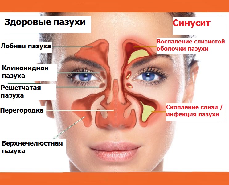
- The paranasal sinuses (sinuses) are located in the bones of the brain and facial skull and communicate with the nasal cavities. They are formed during the growth of the mucosa of the middle nasal passage into the spongy bone tissue. There are several sinuses.
- The frontal sinus is a steam room located in the frontal bone. Frontal sinuses in different people can be developed to varying degrees, in some they are absent. The frontal sinus with the nasal cavity is connected by the fronto-nasal canal, which opens into the anterior part of the lunar fissure in the middle nasal passage.
- The maxillary sinus is located in the body of the upper jaw. This is the largest air cavity of the skull. In the anterior part of the medial wall of the sinus, a nasolacrimal canal passes. The sinus outlet is located behind the nasolacrimal canal at the highest sinus site. The back and bottom of this hole may be an additional hole.
- The lattice labyrinth is a complex multi-chamber cavity.
- The sphenoid sinus is a paired cavity located in the body of the sphenoid bone. The bottom of the sinus forms the arch of the nasopharynx. The hole is in the front wall, connects the sinus with the superior nasal passage. The openings of the optic nerves are located in the upper lateral section.