Diagnosis of gastric and duodenal ulcer, neoplasms (benign, malignant) in modern medicine are very important measures that allow you to control your health and take timely measures when pathologies are detected. Quite a lot of effective research options for the human body have been developed, and instrumental ones that give accurate and detailed information about the patient's condition attract particular attention. In most cases, the diagnosis of gastric ulcer does not present any particular difficulty for doctors, the most important thing is to go to the hospital on time at the first suspicion of a pathology.
Instrumental research
Such techniques are a large and significant section of the examination if a person has a suspected gastrointestinal tract disease. A lot of approaches have been developed. Diagnosis of the stomach can be carried out using an endoscope, x - ray radiation, ultrasound. Electrographic, electrometric approaches are applied. Actual diagnostic methods for the stomach are selected, evaluating the patient's condition and existing complaints. For various diseases, the most suitable option will show more useful information than all others, so a qualified doctor should choose it. The volume of diagnostic measures also depends on the equipment available at the clinic.
The use of a suitable method for the diagnosis of the stomach results in data on the morphology, functional features of a particular tissue site. If the patient is assigned several studies, these are not duplicate ones, set to clarify the information, but necessary to reveal all aspects of the ongoing process of the event. Correct diagnosis of the stomach allows you to determine the relationship of the affected organ and the surrounding tissues, as well as assess the nature of the pathology and its scale.
Attention to the little things!
Before carrying out measures for the diagnosis of the stomach, the doctor explains in detail to the patient how to prepare for the study. It is important to follow all recommendations: this determines how accurate and reliable the information received during the work will be. If the rules established by the doctor are not complied with, there is a chance of obtaining a distorted picture when the present disease cannot be detected, or a diagnosis of stomach diseases will show a violation that does not exist in reality. Many modern methods are very accurate and sensitive, which leads to such a careful attitude.
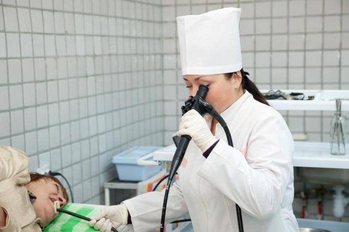
Endoscope in action
One of the most widely used methods for diagnosing stomach diseases is endoscopic examination. As part of the event, the doctor examines the digestive system of the patient from the inside, examining the tissues with which the systems are lined on the inner surface. Thus, it is possible to examine cavities, organs in the form of tubes. A special device is used - an endoscope.
In the diagnosis of stomach diseases , flexible thin tubes are used, supplemented by special optics - they are called endoscopes. The image, a beam of light beams are transmitted via fiber optic cable. Modern stomach diagnostics involves the use of technically advanced equipment, so this technique is completely safe if the event is conducted by a qualified doctor.
Some features: do not rush with treatment
Diagnosis before treatment of the stomach should be carried out as responsibly and in detail as possible in order to collect all the information about the course of the disease. An endoscope can be used when a patient complains of problems with the esophagus and duodenum. This procedure is indispensable before starting a therapeutic course that affects the sigmoid, rectum, colon, stomach. For each of these organs, you need to use a specialized endoscope, designed taking into account human anatomy and physiology. If there is a suspicion of gastric ulcer, diagnosis and treatment usually begin with endoscopy.
The role of this method for detecting abnormalities in the gastrointestinal tract cannot be underestimated, and its importance is growing from year to year, since the latest devices allow not only to visually assess the state of tissues, but also to obtain samples of biological material. On the basis of tissues, a cytological analysis is performed, and structural features and structures of the cells forming the mucous membrane are recognized. In some cases, the doctor prescribes a histochemical, histological examination, materials for which can also be obtained during endoscopy.
Method Features
Using modern equipment, during the study with an endoscope, you can take pictures of the inner surface of the studied organs. For this, special attachments for photographing are used. The event will help document changes in tissue structure.
In some cases, you can record a session on a VCR for more information. This makes it possible to trace the dynamics of the disease, the healing of disorders, if a second examination of the organs is prescribed. This is typical for situations where you need to control how well the ulcer healed, how polyps develop.
Diagnosis and treatment
Using an endoscope, you can not only identify the pathology of the gastrointestinal tract and clarify information about it, but also conduct therapeutic measures. With the help of such a device, polyps of small size are removed, bleeding sites are treated. Affected areas can be sealed, cauterized or laser treated. If ulcers, erosion are detected, the affected organ element is treated with drugs.
To clarify the information you need to use an improved version of the device - a videoscope.
Disease Ultrasound
Ultrasound is a whole range of activities involving scanning, echolocation, echo, sonography. The methodology is based on the fact that waves of a certain frequency, specially directed, focused, can be absorbed, reflected by organs, tissues of the human body due to the different density of these structures. The reflected pulse is converted, recorded and displayed on the screen, from where the data is transferred to the film.
Ultrasound helps to fix the structure of organs, especially position, size, shape. Thus, the stomach, liver, and glands are diagnosed. During the research process, you can find stones, neoplasms, problems with blood vessels, damaged ducts and some other disorders of the normal functioning of internal systems.
Features
To get accurate information, the study is best done in the morning, before this you can not eat. Preparation involves the adoption of drugs that exclude gas formation. The gases accumulated in the intestinal loops do not allow ultrasonic waves to pass into the tissue of the target of the study, which means that the event will not provide accurate information to assess the condition of the patient. Usually, three days before the study, dishes in which the concentration of fiber is increased are completely removed from the diet. Writing down the direction for diagnosis, the doctor tells the patient in detail which products should not be consumed shortly before ultrasound.
If the patient has constipation, flatulence worries in a pronounced form, in addition to the study they drink medicinal carminative decoctions. Dill seeds, coriander, caraway seeds, oats come to the rescue. It will not be superfluous to use activated carbon for three consecutive days. Drink a gram four times a day.
Radiation diagnosis of the stomach
X-rays are one of the accurate studies that allow you to get the most information about the state of the body. Without such an event, the diagnosis of the patient's condition can rarely be called complete, but there are many cases where the x-ray was the only method that showed deviations, pathologies that provoked negative symptoms. Often, such a study can detect factors that threaten a person’s life, while alternative methods are simply not capable of this, no matter how perfect they are in their field.
During the x-ray, the doctor receives information about the digestive tract. Information reflects the shape, location, topography of the mucous membrane, peristalsis. X-ray is absolutely indispensable for suspected ulcers, neoplasms, abnormalities that accompany gallstone disease. You can not do without such a diagnostic method, if the doctor suggests complications - stenosis, varicose veins, penetration. The method is resorted to to identify functional disorders.
When is not so relevant?
A relatively small benefit is an X-ray examination if the patient is diagnosed with gastritis, duodenitis, and colitis. Not too much information can be obtained in the case of cholecystitis. The listed pathologies may not at all affect the images obtained during the diagnosis.
How is it going?
To obtain information during the study, you need to use a special contrast agent - barium sulfate, which is introduced into the body in the form of an aqueous suspension. Such a compound can absorb x-ray radiation, so that the entire route of the tube moving through the digestive system becomes clearly visible in the pictures.
Usually, a study is prescribed in the morning, and a day before the diagnosis it is recommended to eat a little and light food. No special diet is required, but dinner should be eaten small. Recommended porridge, of the preference should be given to tea. Before the diagnosis, you should not smoke, take medications, eat or drink - this will distort information.
Important Features
It is known that the correctness of the result is to some extent determined by the presence of gases in the intestine. This is more characteristic of severe flatulence. Patients with long-term constipation problems may have problems. Under the influence of accumulated gases, the intestinal loops move, put pressure on the stomach, and this prevents a specialized examination. If there is a suspicion of difficulty with an X-ray, an enema is prescribed to the patient two hours before the event.
The accuracy of the results may be affected by mucus and liquid accumulated in the gastrointestinal tract. To prevent such problems, immediately before the diagnosis, the sister flushes the stomach using a probe. For pumping biological fluids, you can use a special volume syringe.
Individuality as a way to success
When conducting a study using x-ray radiation, the doctor always evaluates the characteristics of a particular patient. The procedure is individual, in many respects it is determined by the condition of the patient, and pathology - its location, character, scale. As part of diagnostic measures, fluoroscopy, a review, and radiography are performed. During the procedure, the patient will have to change position several times so that the pictures describe the affected area from all sides.
Nowhere easier
Quite often, patients are prescribed a fairly simple study using an X-ray study. The contrast medium moves through the small, large intestine. Inspection is prescribed not only on the day of the event, but also on the next. When a stool delay is detected or barium moves very slowly, observations last for three days.
If it is necessary to examine the cecum, eight hours before the event, the patient drinks a glass of liquid with barium. This time period is sufficient for the substance to fill the ileum, the appendix. Pictures taken as a result of the event will show the shape and position of the organs. The doctor will be able to evaluate how much they are offset relative to the anatomically correct position.