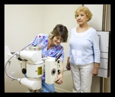
X-ray of the spine is a standard procedure for obtaining images of internal tissues, bones and organs on the film. It is prescribed for many reasons, including for the diagnosis of tumors or damage to bones.
X-ray of the spine can be performed to evaluate any part of the spine:
- The cervical, which consists of seven vertebrae.
- Thoracic, consisting of 12 vertebrae.
- Lumbar: five vertebrae in the lower back.
- The sacrum, which consists of five small fused vertebrae.
They are all about the same size, but differ in structure, coating and articular surfaces. The size of the image is determined taking into account the disease and patient complaints.
X-ray of the spine can be prescribed:
- to identify the causes of pain in the back or neck;
- in case of fracture, arthritis, disc degeneration;
- for the diagnosis of neoplasms;
- with disorders of the spine, such as scoliosis, kyphosis or congenital anomalies.
There may be other reasons prompting the doctor to recommend a spine x-ray.
Before agreeing to a survey, you need to get enough information about it. After all, an x-ray of the spine is not an easy procedure. No special preparations are required, but the patient must know that this is a kind of radiation, and must be aware of where, when and who will take pictures of him.
X-ray of the spine is strictly contraindicated in the first trimester of pregnancy, it is allowed only in emergency situations, when a woman's health is more important than the condition of the fetus. This is due to the fact that the fetus has not yet formed and radiation exposure will adversely affect its development.
Before the procedure:
- The doctor must explain the essence of the procedure and answer questions that the patient may have.
- As a rule, there is no preliminary preparation, for example, a diet or taking any medications is not required.
- You must inform your doctor if you expect a pregnancy.
- In addition, a recent barium x-ray of the esophagus may interfere with the quality of the low back image.
As a rule, an x-ray of the spine or the entire back takes place in this way:
- You will be asked to remove jewelry, hairpins, glasses, hearing aids and other metal objects that interfere with the procedure.
- If it will be necessary to remove items of clothing, you will be provided with a special bathrobe.
- You will need to take the appropriate position to get the right shot.
- Parts of the body that are not removed are covered with a lead apron (shield) to avoid exposure to x-rays.
- If the procedure is carried out to determine the injury, special attention should be paid to measures to prevent further damage. For example, an x-ray of the cervical spine may require a neck brace.
- Sometimes you need to take pictures in several different positions. It is imperative to stay in place while the picture is taken, as any movement can lead to image distortion.
- The x-ray beam is focused.
- The doctor for the time of the beam beam goes into the protective room.

An x-ray of the spine does not cause pain, but manipulation of various parts of the body can cause discomfort or pain, especially in the case of recent trauma or invasive procedures such as surgery. The doctor should use all possible measures to ensure comfort, complete the procedure as quickly as possible and minimize pain.
Diagnostics of the spine, back, or neck can also be done using myelography (myelogram), computed tomography (CT), magnetic resonance imaging (MRI), or bone scan.