A symptom such as a headache is familiar to everyone. Often the cause of its appearance are vascular diseases. In most cases, they develop in middle and old age. Due to insufficient blood supply to the brain , signs of ischemia develop. These include: memory impairment, decreased performance, sleep disturbance. To diagnose ischemia, Doppler imaging of the vessels of the brain and neck (USDG) is performed. Among them are large arteries and veins, originating from the aortic arch. Currently, ultrasound scan is performed in almost all clinics. This study is not harmful to health and painless.
What is vascular dopplerography?
Doppler ultrasound (Doppler ultrasound) is performed to examine veins or arteries. Most often, thanks to this method, I diagnose vascular pathologies of the lower and upper extremities, as well as the head and neck. This study can be performed in patients of any age. There are practically no contraindications to dopplerography. It can be administered to infants and pregnant women. Dopplerography of the vessels of the brain and neck is an instrumental method of research based on determining the speed of blood flow. Thanks to this diagnostic procedure, it is possible to identify how affected the arteries and veins are, the degree of narrowing of their lumen.
Dopplerography: the essence of the method
When prescribing this diagnostic method, patients are interested in: "What is dopplerography?" You should know that this procedure is necessary to assess the condition of the vessels. It is carried out in conjunction with an ultrasound scan (USDG). Using dopplerography, you can find out:
- Speed and direction of blood flow. Often with pathologies of arteries and veins, its slowdown or acceleration, turbulence is observed.
- The condition of the vessels. Thanks to the ultrasound scan, obstruction of the lumen of arteries or veins with atherosclerotic plaques, a thrombus, etc. is determined. In addition, this method allows you to determine the weakness of the valve apparatus of the vessels.
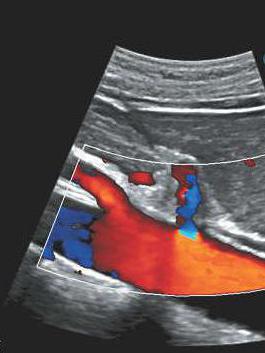
The essence of the study is to scan the arterial or venous system. The image appears on the monitor. There are duplex and triplex scanning of blood vessels. In the first option, it is possible to assess the condition of the endothelium and the lumen of the arteries of the head and neck. Triplex scanning is more complex. With its help, a color image of the vascular system is obtained. This improves visualization, and also makes it possible to assess the state of blood flow.
Doppler methods
Doppler ultrasound ( vascular dopplerography ) of the head and neck is performed in several ways. Each of them is effective in the diagnosis of certain diseases. The choice of dopplerography method depends on which part of the vascular bed needs to be examined. Thus, allocate:
- Carotid Ultrasonography. In this case, using a special device, the vessels in the neck are examined. This method of visualization is the most common. It is used for suspected neurological diseases associated with malnutrition of the brain. The vessels of the neck are located superficially, the largest of them are the carotid arteries. Carotid ultrasonography is indicated for dizziness, fainting, decreased sleep and memory, and neurological symptoms.
- Transcranial duplex scanning. The sensor is located in the occipital, temporal and temporal areas. In this way, dopplerography of the head is performed. Thanks to this method, the anterior, posterior, middle and vertebral arteries are examined. Also, in a similar way, you can assess the state of the direct sinus, veins of Galen and Rosenthal.
- Transorbital scan. The sensor is mounted on the upper eyelid. Using this access, blood flow in the supra-lateral and orbital arteries is evaluated.
Indications for Dopplerography
Dopplerography of the vessels of the brain and neck is performed with suspected circulatory disorders. Chronic ischemia is manifested by symptoms such as impaired memory, sleep, mental and nervous disorders. In acute circulatory disorders, convulsive syndrome, loss of consciousness, paresis or paralysis of the extremities are possible. The following indications are distinguished for conducting ultrasound of the head and neck arteries:
- Transient ischemic attack.
- Stroke.
- Encephalopathy.
- Condition after acute cerebrovascular accident.
- Suspicion of aneurysm or vascular malformation.
- Dizziness and fainting of unknown etiology.
- Osteochondrosis of the cervical spine.
- Arterial hypertension.
- Visual impairment: hemianopsia, diplopia, the appearance of cattle.
Dopplerography during pregnancy is not contraindicated. In some cases, this research method is prescribed if there is a suspicion of a violation of placental circulation. Doppler ultrasound of the veins and arteries of the brain and neck of pregnant women is carried out in the presence of the above pathologies.
In what cases is dopplerography contraindicated?
The main advantage of dopplerography is the absence of contraindications to the study. In fact, USDG is a completely harmless procedure. It can be carried out at any age, even in the presence of chronic diseases. Nevertheless, there are cases when Dopplerography is not recommended. Relative contraindications include severe conditions in which it is difficult for the patient to be in a horizontal position. This can be observed with attacks of bronchial obstruction (COPD, asthma), acute heart and respiratory failure.
Preparation for Dopplerography of the vessels of the head and neck
Dopplerography of the vessels of the brain and neck does not require special preparation. However, before conducting this study, laboratory diagnostics are performed. When a patient complains of headache, sleep disturbance, vision, memory impairment, tests are first prescribed. Among them - UAC, OAM, blood biochemistry, coagulogram. If laboratory data do not indicate the cause of the appearance of such symptoms, then it is necessary to perform an ultrasound scan of the vessels of the neck and head. Before conducting Doppler, you must remove the jewelry. The diagnostic method is not accompanied by radiation exposure to the body. If dopplerography is required during pregnancy, then it should be explained to the woman that this procedure is not dangerous to the fetus.
Survey technique
Regardless of the choice of access, the patient's position is lying on his back. When performing carotid ultrasonography, it is necessary to turn the head to the right or left and slightly tilt it back. The sensor is installed in the area of large and medium vessels. Before starting the study, the surface of the neck is smeared with acoustic gel, which is necessary to slide the device and improve visualization. After that, the device is moved over the skin, while the image of arteries and veins appears on the screen.
With transcranial duplex and triplex scanning, the vessels located in the cranial cavity and outside it are examined. For this patient, they are asked to take a sitting position. To assess the condition of the cerebral arteries, the sensor is installed first anteriorly, and then - above and behind the auricle. The study of the direct sinus, veins of Rosenthal and Galen, parabasilar and cerebellar vessels is possible when moving the device to the occipital region. There are two ways to visualize the vertebral artery : using transcranial scanning or carotid sonography.
The study of the vessels of the organs of vision is carried out in the supine position. In this case, the eyelids of the patient should be closed. In addition, a siphon of the internal carotid artery is visualized using transorbital access.
Doppler results of vessels of the head and neck
After the study, it is necessary to evaluate the results and compare the data with the norm. Carotid ultrasonography is performed to assess the shape of blood vessels, their location, thickness and condition of the lumen. The diastolic and systolic peak blood flow velocities in the carotid arteries are also determined. Normally, it should not exceed 0.9. If this indicator is close to 1.5, then the narrowing of the lumen of the vessel is about 50%. With complete obstruction of the artery, the peak velocity is not determined (equal to 0). This condition occurs with thrombosis or embolism of the vessel and can lead to death. The narrowing of the lumen is observed with atherosclerosis. This disease is common among older people and often causes the development of discirculatory encephalopathy, as well as acute disturbance of cerebral circulation.
When duplex or triplex color scanning, in addition to the above indicators, possible changes in blood flow (speed, direction) are evaluated. It is also possible to visualize the degree of filling of the vessels. Information on the weakness of venous valves can be obtained during functional tests (ask the patient to take a sip, change the position of the body).
USDG of the vessels of the head and neck: the price of the procedure
Dopplerography is performed in most clinics that have a department or functional diagnostic rooms . Before you come to the examination, you must pre-register for ultrasound of the vessels of the head and neck. The price of this procedure will depend on the choice of clinic. On average, it is 2300-3500 rubles. It is advisable to have a referral for research. In some clinics, dopplerography of the vessels of the neck and head is done both individually and together. The price depends on it. The cost of ultrasound of the vessels of the neck (or head) ranges from 1100 to 1800 rubles.
Doctors reviews about dopplerography
According to doctors, vascular dopplerography is the standard in the diagnosis of brain diseases. The advantages of this method are its safety and high information content. In addition, there are no contraindications for ultrasound. SPB (St. Petersburg) is a city in which there are many clinics with equipped rooms for radiation diagnostics. Dopplerography of the vessels of the head and neck can be done in medical centers: Lakhta, MEDEM, Energo, Longevity, etc.