The skeleton of an adult is approximately 206 bones. Each of them has its own structure, location and function. Some bones help to move, others protect our organs and tissues from mechanical damage, while others provide the opportunity to perform actions such as chewing, swallowing and, of course, speaking. It is these functions that the hyoid bone and the muscles that attach to it perform. Despite its very small size, this bone is very important. Injuries associated with her fracture are extremely dangerous, they often end in death.
Anatomical structure
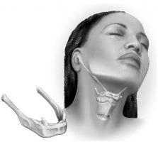
The hyoid bone is located directly under the body of the tongue. She can be felt only by thin people. Its dimensions are relatively small, but it is involved in the performance of very important functions. Together with the muscles that connect with it, it helps to carry out processes such as chewing and swallowing. In addition, human speech would have been impossible without it. So it’s impossible to overestimate the value of this bone. The structure of the hyoid bone is simple. It is conditionally divided into body, large and small horns. It connects to the rest of the bones using joints and ligaments. The body of the hyoid bone has the shape of an uneven plate, slightly convex in front. It has vertical and transverse ridges. The edges are also heterogeneous: the upper one is pointed, and the lower one, on the contrary, is slightly thickened. From the sides, the body connects with the help of the articular surfaces of the cartilage with large horns. They move backward. Large horns are much longer and thinner than the body. At the ends, they can detect thickenings. From the place where the large horn connects to the body, small horns depart. As a rule, they consist of bone tissue, but in some cases remain cartilaginous. They connect to the body also through the joint. The ends of the small horns are enclosed in the stylohyoid ligament. Sometimes it contains one, less often several rather small bones.
Hyoid fracture and symptoms of pharyngeal damage
Fractures and damage to the hyoid bone are quite rare. As a rule, this occurs as a result of a blunt trauma of the submandibular region. At the same time, a rather strong mechanical effect should be exerted on this site. In some cases, a fracture can cause strangulation. This also happens when hanging. Fresh small fracture makes itself felt quite obvious symptoms. First of all, it is severe pain in the upper front of the neck when swallowing or chewing. Also in the area of the hyoid bone, a small hematoma will be noticeable. When palpation is felt mobility and crepitus debris.
In severe trauma of the hyoid bone, mucosal rupture occurs. This is accompanied by fairly severe bleeding from the mouth. It occurs due to damage to the branches of the lingual or thyroid artery. Often such an injury is fatal. First aid for fractures of this nature is very difficult and not always effective.
We can say that all injuries in which the hyoid bone is involved (you can see a photo of its location in the article) are very dangerous for human health and even human life.
First aid
First aid for fracture of the hyoid bone should be carried out quickly. When there is excessive bleeding from the mouth, the blood coagulation process must be activated. This can be done by tamponade or by applying cold. If possible, then you need to try to bandage the external carotid artery. After the injury, the first hours are the most dangerous. Making any predictions is very difficult due to the risk of developing asphyxiation. If the pharynx ruptures, too much blood loss is possible. Unfortunately, death often occurs before the ambulance is called.
Indeed, it is extremely difficult to help a person when the hyoid bone is broken and the mucous membrane is torn. If there are all signs of asphyxiation, then the best thing to do is to intubate the trachea and then swallow the throat to reduce blood loss. After these complex manipulations, the victim must be taken to the hospital as soon as possible.
Treatment
The treatment of injuries associated with a fracture of the hyoid bone is to immobilize and completely eliminate all displacements of the fragments. This can be achieved by the palpation method both from the side of the oral cavity, and, of course, from the outside. Immobilization of the head and, very importantly, the neck is carried out using a securely fixing corset. In severe cases, when the hyoid bone is badly damaged, plaster is placed on the shoulders and neck. But in practice, most often, maintaining bone fragments in the correct position is achieved only with surgical reposition. Often such injuries entail a number of complications, so treatment should be as effective as possible.
Hyoid muscle
All muscles that are attached at one end to the hyoid bone are conventionally divided into two groups: sublingual and sublingual. They differ from each other in position and, accordingly, in functions. The suprahyoid muscles include:
- double-abdominal;
- maxillohyoid;
- awl-sublingual;
- chin-hyoid muscle.
All of them are located above the hyoid bone and are directly attached to it. The biceps muscle consists of the anterior and posterior abdomen, which are connected by tendons. It is closely interconnected with another group of fibers. The posterior abdomen in its upper part is attached to the temporal bone. Going down, the latter is adjacent to the stylohyoid muscle and passes into the intermediate tendon. It covers the fixation loop of the body and the large horn of the hyoid bone. But before that penetrates the stylohyoid muscle, which has a fusiform shape. Another group of fibers departs from the lower jaw from its inner surface. The maxillary hyoid muscle is flat and wide. The bundles of its fibers are located transversely, they are directed towards and grow together, forming a tendon suture. On the side of the midline of the maxillo-hyoid begins the chin-hyoid muscle.
Functions of the Sublingual Muscles
A group of suprahyoid muscles performs one common function. They allow the hyoid bone to move up, down and to the sides. This helps a person perform complex tasks such as swallowing and chewing. Thus, it can be said that suprahyoid muscles are involved in the digestive and respiratory functions, albeit indirectly. Also, this group of muscle fibers, raising the hyoid bone along with the larynx and lowering the lower jaw, contributes to the process of speech formation.
Sublingual muscles
The sublingual muscles include the following: sternum-hyoid, scapular-hyoid, sternum-thyroid muscle. They are also attached to the hyoid bone, but are located below it. So, the
scapular-hyoid muscle begins in the upper part of the scapula. It has two large abdomen, which are separated by an intermediate tendon. The
sternum -hyoid muscle with its lower end is attached to the
handle of the sternum. It, like the scapular-hyoid fibers, joins the hyoid bone with its upper part. The third muscle group - the sternum-thyroid - lies in front of the thyroid gland and trachea.
Sublingual Muscle Functions
Sublingual muscles, acting as a group, pull the hyoid bone together with the larynx down. But each of them at the same time performs one specific function. For example, the sternum-thyroid muscle selectively moves the thyroid cartilage down. But no less important is another function of the sublingual muscles. Reducing, they reliably strengthen the position of the hyoid bone, to which the suprahyoid muscle group is attached, while lowering the lower jaw.