In all world cultures, the function of reproduction, procreation, is considered one of the main. The male and female reproductive system has a different structure, but performs one task: to form germ cells - gametes, at the merger of which, at the time of fertilization, the development of the future human body will become possible. This article is devoted to the study of the structure and function of the female reproductive system.
General characteristics of the reproductive organs of women
The female reproductive system includes the external and internal genital organs, which are also called reproductive (reproducing).
The external genitalia, called the vulva, are visually expressed sufficiently - this is the pubis, the labia minora and the labia minora, the clitoris and the entrance to the vagina (vagina), which is closed by an elastic hymen called the virgin. We will study the external organs of the female reproductive system in more detail.
Pubic structure
The lower abdomen at the level of the pubic (pubic bone) forms the pubis. The bone itself, with an anatomically correct position, hangs over the entrance to the vagina and looks like an arch. Outwardly, the pubis has a roll-like shape, forming an elevation. A layer of fat forms under his skin. Outside, puberty forms on it during puberty . It has a distinct horizontal border. If an excess amount of androgens, male sex hormones, is produced in a woman’s body, her hairline increases and rises at an acute angle to the navel. Pathology of pubic hair is a sign of sexual development.
Labia minora and labia minora
From the pubis to the anus, there are two folds of skin - the labia majora, which has an external hairline and a layer of subcutaneous fatty tissue. In their connective tissue are the ducts of the Bartholin gland. It secrets a fluid that moisturizes the female genital organs. In violation of hygiene, harmful microorganisms penetrate into the tissues of the gland and cause inflammation in the form of painful seals.
Under the large are the labia minora, densely braided by blood vessels and nerves. In their upper part is an organ homologous to the male penis - the clitoris. Its growth is inhibited by hormones of the female reproductive system - estrogens. The clitoris contains a large number of nerves and blood vessels, which means it has a high sensitivity. If the size of the clitoris is greatly increased in a girl or woman, this can be a clear sign of hormonal pathology.
Vaginal entrance
The vulva, in addition to the pubis, the labia minora, the labia minora, the clitoris, includes the entrance to the vagina. At a distance of 2 centimeters from it inland is the hymen. It consists of connective tissue and has several holes through which blood flows during menstruation.
Female reproductive organs
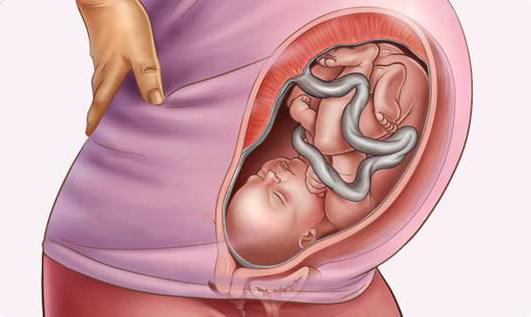
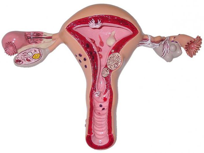
These include the vagina (vagina), the uterus, ovaries, and the uterine (fallopian) tubes. All of them are located in the pelvic cavity. Their functions are the maturation and entry of fertilized female reproductive gamete-egg cells into the uterine cavity. The embryo will develop in it from the zygote.
Vaginal structure
The vagina is an elastic tube made up of muscle and connective tissue. It is located from the genital fissure towards the uterus and has a length of 8 to 10 cm. Located in the pelvis, the vagina enters the cervix. It has anterior and posterior walls, as well as an arch - the upper section of the vagina. The back of the vagina is deeper than the front.
The vagina is located at an angle of 90 degrees to the surface of the uterus. Thus, the internal female genital organs, which include the vagina, are densely braided by arterial and venous vessels, as well as nerve fibers. The vagina is separated by a thin connective tissue wall from the bladder. It is called the vesicovaginal septum. The lower part of the vaginal wall at the back is separated from the lower part of the large intestine by the perineal body.
Cervix : structure and function
The vagina enters the canal, called the cervical, and the junction itself is the external pharynx. Its shape is different in women giving birth and not giving birth: if the pharynx is point-oval - the uterus did not bear the fetus, and the appearance of the gap is characteristic of the women giving birth. The uterus itself is an unpaired hollow muscle organ, consisting of the body and neck and located in the pelvis. Considering the structure of the female reproductive system and its function, it becomes clear that she is responsible for the formation and development of the embryo, as well as for the process of expulsion of the fetus as a result of labor. Let us return to the structure of its lower part - the neck. It is connected to the upper part of the vagina and has the shape of a cone (for nulliparous) or a cylinder. The vaginal area of the cervix has a length of up to three centimeters, and is also anatomically divided into the front and rear lips. The cervix and pharynx transform with the age of the woman.
Inside the cervix is the cervical canal, ending with an internal pharynx. It is lined with secretory glands that secrete mucus. If its secretion is disturbed, clogging and cyst formation may occur. Mucus has bactericidal properties and prevents infection of the uterine cavity. 4-6 days before the ovum leaves the ovary, the mucus becomes less concentrated, so sperm can easily penetrate through it into the uterus, and from there into the fallopian tubes.
After ovulation, the cervical secretion increases its concentration, and its pH decreases from neutral to acidic. The pregnant uterus is completely covered by a clot of cervical mucus in the neck. During the menstrual period, the cervical canal slightly opens so that the torn endometrial layer can exit. This may be accompanied by aching pains in the lower abdomen. During labor, the cervical canal can open up to 10 cm in diameter. This contributes to the birth of a child.
Among the most common diseases of the cervix can be called its erosion. It appears as a result of damage to the mucous layer caused by infections or injuries (abortions complicated by childbirth). Timely undetected and untreated erosion can cause inflammatory processes and even cancer.
Fallopian tubes
The fallopian tubes, also called oviducts or fallopian tubes, are 2 elastic tubes located in the abdominal cavity and entering the bottom of the uterus. The free edge of the oviduct has fringes (fimbriae). Their beating ensures the advancement of an egg that leaves the ovary into the lumen of the tube itself. The length of each oviduct is from 10 to 12 cm. It is divided into sections: a funnel that has an extension and is equipped with fimbriae, an ampoule, an isthmus, a part of the canal entering the uterine wall. For the normal development of pregnancy, a condition such as complete patency of the oviducts is necessary, otherwise the woman expects infertility. The most common pathologies of the fallopian tubes, such as adhesions, salpingitis and hydrosalpinx.
All of these diseases cause tube infertility. They are complications of chlamydia, gonorrhea, trichomoniasis, genital herpes, causing narrowing of the lumen of the fallopian tubes. Frequent abortions can provoke the appearance of adhesions that are located across the tube. Hormonal disorders cause a decrease in the mobility of the ciliary epithelium lining the oviducts, which leads to a deterioration in the motor properties of the egg.
The most dangerous complication resulting from tubal pathologies is an ectopic pregnancy. In this case, the zygote stops in the oviduct without reaching the uterus. It begins to crumble and grow, stretching the tube wall, which, in the end, bursts. As a result, severe internal bleeding occurs that threatens life.
Ovaries in women
They are a paired sex gland and have a mass of 6-8 grams. The ovaries are glands of mixed secretion. The production of sex hormones - estrogens, controlled by the pituitary gland and hypothalamus - is an intra-secretory function. Like glands of external secretion, they form germ cells - gametes, called ova. The biochemical composition and mechanism of action of estrogen will be studied by us later. Let us return to the structure of female gonads - ovaries. It should be borne in mind that the structure of the female reproductive system (as well as the male) is directly related to the urinary system.
It is from the mesonephros (primary kidney) that the stroma of female gonads develops. The oocyte precursors are oogonium, formed from mesenchyme. The ovary has a protein coat, and under it two layers: cortical and cerebral. The first layer contains follicles, which, ripening, form ovocytes of the first and second order, and then mature eggs. The brain substance of the gland consists of connective tissue and performs a supporting and trophic function. It is in the ovaries that ovogenesis occurs - the process of reproduction, growth, and maturation of female reproductive gametes - eggs.
The specificity of the hormonal background in a woman
The structure of the reproductive system of the female and male individuals is controlled by special biologically active substances - hormones. They are produced by the sex glands: testicles in men and ovaries in women. Entering the bloodstream, they address both the development of reproductive organs and the formation of secondary sexual characteristics: body hair growth, mammary gland development, pitch and timbre of the voice. The development of the female reproductive system occurs under the influence of estradiol and its derivatives: estriol and estrone. They are produced by special ovarian cells - follicles. Female hormones - estrogens lead to an increase in the volume and size of the uterus, as well as muscle contractions of the fallopian tubes and the uterus itself, that is, the reproductive organ is prepared for the adoption of the zygote.
The corpus luteum of the uterus produces progesterone - a hormone that stimulates the development of a child's place - the placenta, as well as an increase in the glandular epithelium of the mammary glands during pregnancy. Violation of the hormonal background of the female body leads to diseases such as uterine fibroids, endometriosis, polycystic.
Anatomical features of the female uterus
The reproductive system of the female body incorporates an organ that is unique in structure and function. It is located in the pelvic cavity between the bladder and rectum and has a cavity. This organ is called the uterus. To understand the mechanism of fertilization, we recall that the genitals - the ovaries in women, are associated with the fallopian tubes. The egg, entering the oviduct, then enters the uterus, which serves as the organ responsible for the development of the embryo (embryogenesis). It consists of three parts: the neck, which has been studied previously, as well as the body and bottom. The body of the uterus looks like an inverted pear, in the expanded part of which two fallopian tubes enter.
The reproductive organ is covered with a connective tissue membrane and has two layers: muscle (myometrium) and mucous (endometrium). The latter is built from squamous and cylindrical epithelium cells. The endometrium changes the thickness of its layer: during ovulation, it thickens, and if fertilization does not occur, this layer is rejected along with a portion of blood from the walls of the uterus - menstruation occurs. During pregnancy, the volume and size of the uterus increases greatly (about 8-10 times). In the pelvic cavity, the uterus is suspended on three ligaments and braided by a dense network of nerves and blood vessels. Its main function is the development and nutrition of the embryo and fetus until the time of physiological birth.
Uterine pathology
The structure of the reproductive system of a female individual may not always be ideal and correctly functioning. One of the pathologies of the reproductive system associated with the structure of the genital organ may be a bicornuate uterus. It has two bodies, each of which is associated with one oviduct. If the pathology of the female reproductive system concerns the structure of the endometrium, they speak of hypoplasia and aplasia of the uterus. The consequence of all the above pathologies is the termination of pregnancy or infertility.
In this article, the anatomical and physiological features of the female reproductive system were studied.