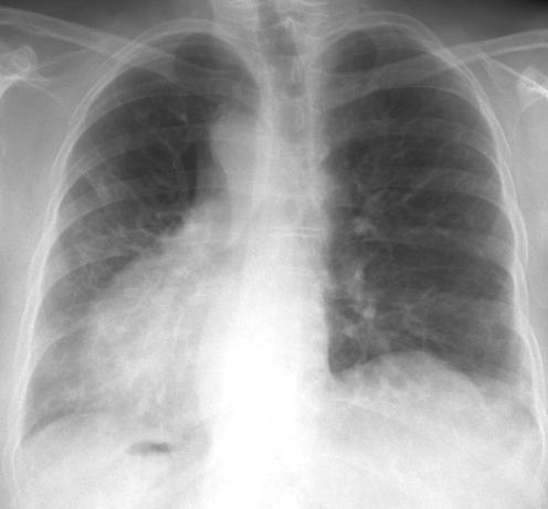
The human lungs are one of the most important organs, without which its existence is impossible. Breathing seems to us so natural, but in fact, during it, complex processes occur in our body that ensure our vital functions. To better understand them, you need to know the structure of the lungs.
In the process of breathing, air passes through two bronchi, which have a different structure. The left one is longer than the right one, but narrower than it, therefore most often the foreign body enters the respiratory system through the right bronchus. These organs have branches. Upon entry into the lung, the right branch for 3, and the left for 2 lobes, which corresponds to the number of lobes of the lungs.
The structure of the lungs is quite complex, because inside them the bronchi branch into many small segmental bronchi. In turn, they pass into lobular bronchi entering the lobules of the lungs. It is difficult to imagine what the structure of the lungs is, not knowing how many lobular bronchi are in them (there are about 1000 of them). Intralobular bronchi have up to 18 branches (terminal bronchioles) that do not have cartilage in their walls. These terminal bronchioles form the structural component of the lungs - the acinus.
The structure of the lungs is easier to learn, understanding what acinus is. This structural unit is a collection of alveoli (derivatives of respiratory bronchioles). Their walls are a material substrate for gas exchange, and the area during a full breath can reach 100 sq.m. The greatest stretching of their respiratory surface occurs during physical exertion.
The bronchopulmonary segment is the part of the pulmonary lobe that is ventilated by 3-order bronchi branching from the lobar bronchus. Each of them has a separate broncho-vascular pedicle (artery and bronchus). The segmental structure of the lungs was revealed during the development of the level of medicine and surgery. In the right lung there are 10 segments, and in the left - 8. Due to the fact that the division of the lungs into bronchopulmonary segments was established, it became possible to remove the affected areas of this organ with the maximum preservation of its healthy parts.
In this organ, it is customary to distinguish the following surfaces: mediastinal, diaphragmatic, costal. In the mediastinal there are so-called “gates”. Through them, the bronchi, arteries and nerves enter the lungs, and the lymphatic vessels and pulmonary veins exit . All these formations make up the so-called “root of the lung”.
The lungs are separated by grooves of various depths and lengths. They divide the tissues to the gates of the lungs themselves. There are 3 lobes of the right lung (lower, upper, middle) and 2 left (lower, upper). The lower lobes are the largest.
The structure of the lungs will be incomplete without taking into account the visceral pleura sheets, which cover each lung and the root area and form a “parietal leaf” lining the walls of the chest cavity. Between them there is a slit-like cavity, part of which is called sinuses (located between the parietal leaves). The largest pleural sinus is considered to be the costo-diaphragmatic (the edge of the lung falls into it when inhaling).
The structure of the lungs explains the processes that occur in them during breathing. In this body, there are 2 systems of blood vessels: a small circle (consists of veins and arteries involved in gas exchange), a large circle of blood circulation (consists of bronchial arteries and veins that deliver arterial blood to ensure metabolism and support the vital functions of the lungs). By the nature of their branching, the pulmonary veins are similar to arteries, but differ in their inconstancy. Their source is the capillary network of lobules, interlobular connective tissues, small bronchi and visceral pleura. Interlobular veins merging from each other are formed from capillary networks. Of these, larger veins form, passing near the bronchi. Two veins are formed from the lobar and segmental veins in each lung: the lower and upper veins (their sizes vary greatly). They separately fall into the left atrium.
The number of bronchial arteries is variable. It ranges from 2 to 6. In 50% of cases, a person has 4 bronchial arteries that run evenly to the left and right main bronchi. They are not exclusively the arteries of the bronchi, because they give branches to different organs of the mediastinum. The beginning of the right arteries is located in the tissue behind the esophagus and in front or under the trachea (between the lymph nodes). The left arteries are located in the tissue below the trachea and under the aortic arch. Inside the lung, the arteries are in the fiber along the bronchi and, branching, play a direct role in the blood supply to the rest of its parts and pleura. In respiratory bronchioles, they lose their independent significance and pass into the capillary system.
All blood vessels of the lungs are connected to each other. In addition to the general capillary network, extraorganic and intraorganic anastomoses are distinguished, connecting both circles of blood circulation.
The lymphatic system consists of the initial capillary networks, the plexus of the lymphatic vessels inside the body, the discharge vessels, extrapulmonary and intrapulmonary lymph nodes. Distinguish between superficial and deep lymphatic vessels.
The source of lung innervation is the nerve plexuses and trunks of the mediastinum, formed by the branches of the sympathetic, vagus, spinal and phrenic nerves.