Nerve fiber is a process of a neuron that is covered with a glial membrane. What is it for? What functions does it perform? How is it arranged? You will learn about this from the article.
Classification
Fibers of the nervous system have a different structure. According to their structure, they can be one of two types. So, emit myelin and myelin fibers. The first consists of a process of the cell, which is located in the center of the structure. It is called an axon (axial cylinder). This process is surrounded by a myelin sheath. Given the nature of the intensity of the functional load, the formation of nerve fibers of one type or another occurs. The structure of structures directly depends on the department in which they are located. For example, myelin nerve fibers are located in the somatic department of the nervous system , and bezmyelinovye - in the vegetative one. It should be noted that the process of formation of those and other structures takes place according to a similar scheme.
How does a thin nerve fiber appear?
Consider the process in more detail. At the stage of the formation of bezmyelin-type structures, the axon deepens into a cord consisting of lemmocytes, in which the cytolemma begins to bend and cover the appendix according to the principle of coupling. At the same time, the edges are closed above the axon, and a duplication of the cell membrane is formed, which is called "mesaxone". The lemmocytes in the neighborhood form simple contacts with their cytolemma. Due to poor isolation, non-myelin fibers are able to pass a nerve impulse both in the region of mesaxone and in the area of contacts between lemmocytes. As a result, it moves from one fiber to another.
The formation of thick structures
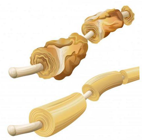
The myelin-type nerve fiber is significantly thicker than the myelin-free. In the process of forming shells, they are the same. Nevertheless, the accelerated growth of neurons in the somatic department, which is associated with the development of the whole organism, contributes to the extension of mesaxons. After that, lemmocytes several times wrap around axons. As a result, layers of a concentric type are formed, and the nucleus with the cytoplasm is moved to the last turn, which is the outer sheath of the fiber (non-acceptable). The inner layer consists of mesaxone entwined several times, and is called myelin. Over time, the number of turns and the size of mesaxone gradually increase. This is due to the passage of the myelination process during the growth of axons and lemocytes. Each next round is wider than the previous one. The widest is the one that contains the cytoplasm with the nucleus of a lemmocyte. In addition, the thickness of myelin varies along the entire length of the fiber. In those places where the lemmocytes come into contact with each other, the layering disappears. Only the outer layers come into contact, which include the cytoplasm and the nucleus. Such places are formed due to the lack of myelin, thinning of the fiber and are called nodal interceptions.
The growth of structures in the central nervous system
Myelination in the system proceeds as a result of grasping by the processes of axon oligodendrocytes. Myelin consists of a lipid base and, when reacted with oxides, acquires a dark color. The remaining components of the membrane and its gaps remain bright. Such strips found are called myelin notches. They correspond to insignificant layers in the cytoplasm of a lemmocyte. And in the axon cytoplasm there are neurofibrils and mitochondria located longitudinally. The largest number of them is closer to intercepts and in fiber end devices. Axon cytolemma (axolemma) contributes to a nerve impulse. It manifests itself in a wave of its depolarization. In the case when neurite is represented as an axial cylinder, it does not contain granules of a basophilic substance.
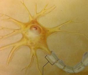
Structure
Myelin nerve fibers are composed of:
- Axon, which is located in the center.
- Myelin sheath. It covers the axial cylinder.
- Schwann shell.
The axial cylinder contains neurofibrils. The myelin sheath consists of many lipoid substances that form myelin. This compound is of great importance in the activity of the central nervous system. In particular, the speed with which the excitation is carried out along nerve fibers depends on it. A shell formed by the compound covers the axon in such a way that gaps are formed that are called Ranvier intercepts. In their area, the axial cylinder is in contact with the Schwann shell. A fiber segment is its gap that lies between two Ranvier intercepts. In it, you can consider the core of the Schwann shell. It is located approximately in the center of the segment. It is surrounded by the protoplasm of the Schwann cell with the content of myelin in the loops. At Ranvier intercept intervals, the myelin sheath is not uniform. It contains oblique notches Schmidt-Lantermann. The cells of the Schwann membrane begin to develop from the ectoderm. Under them is the axon of the fiber of the peripheral nervous system, due to which they can be called glial cells. The nerve fiber in the central system is devoid of Schwann sheath. Instead, there are elements of oligodendroglia. The non-myelin fiber contains only the axon and Schwann sheath.
Function
The main task that the nerve fiber performs is innervation. This process is of two types: pulse and pulseless. In the first case, transmission occurs due to electrolyte and neurotransmitter mechanisms. Myelin plays the main role in innervation; therefore, the rate of this process is much higher in myelin fibers than in bezmyelin. The pulseless type process occurs by the axoplasm current passing through special axon microtubules that contain trophogens (substances that have a trophic effect).