The skeleton of a person’s hand can be divided into 4 sections. The upper is the belt of the upper limb. This includes the scapula and collarbone. Next comes the anatomical shoulder itself, i.e., the division of the humerus. The next section is the forearm, consisting of the ulna and radius. The latter is the bones of the brush. The skeleton of the left hand is a mirror image of the skeleton of the right.
Section Overview
Consider the skeleton of the hand for each section. The shoulder blade and collarbone are connected to each other, and the ball joint connects them to the humerus. But not only the humerus joins them. They serve as a fastening place for the muscles that are responsible for the movement of the arm.
Next is the humerus itself. To her through the elbow joint, the beam and elbow are attached. The latter are movable relative to each other. With the position of the hand, when the palm looks inward, these bones are parallel, but it is worth turning the palm forward, as they move and cross.
The skeleton of the hand is the most complex structure . The composition includes 27 bones. These elements are further divided into several groups: the wrist, metacarpus and phalanges of the fingers, connecting through interphalangeal joints. It is the complexity of this apparatus that allows the hand to be so versatile and skillful. With its help, you can do rough work with mechanical operations, but it also allows you to perform subtle precise movements.
The detailed structure of the shoulder girdle
The skeleton of the arm in the shoulder girdle is represented by the scapula and collarbone. It is the area of their placement and connection with the humerus that is called the shoulder in everyday life. However, the anatomical shoulder is the humerus, and these elements make up the belt of the upper limb. But, considering the skeleton of the human hand, the structure must be studied together with the shoulder girdle, which significantly affects functionality.
Shovel
Shoulder - flat bone on the back. It has a triangular shape with upper, lateral and medial edges and lower, upper and lateral corners. It is the thickened lateral angle that is provided with the articular cavity, where the scapula joins with the head of the humerus located in the next section. A little above the cavity is the neck of the shoulder blade, which looks like a narrowed place. The joint cavity is also surrounded by tubercles - subarticular and superarticular.
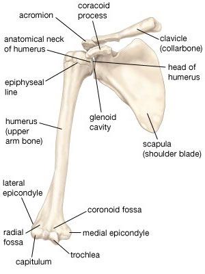
The scapula itself has a slightly concave surface - the scapular fossa - in the region of the ribs from the side of the chest. But on the back surface there is an awn that runs along the scapula from the inner edge to the outer corner. On the sides of the spine, the supraspinatal and infraspinatous fossae are distinguished, where muscles with the same names are attached. Outside, this spine passes into the shoulder process located above the shoulder joint, called the acromion. The scapula is also equipped with a coracoid process, facing forward and serving to attach the ligaments and muscles.
Collarbone
The clavicle is a tubular bone, curved S-shaped. Has a horizontal position, goes to the upper front of the chest near the neck. The medial sternal end is attached to the sternum, and the acromial lateral is connected to the scapula. Mounting is also done with muscles and ligaments, which causes the presence of roughness on the lower surface, namely the line and tubercle.
Shoulder structure
Behind the shoulder girdle is directly the skeleton of the human hand. The shoulder is formed precisely by the humerus. This is a tubular bone, rounded in cross section from the upper side and trihedral closer to the bottom. The upper end is crowned with a head in the form of a hemisphere, which is turned towards the scapula. There is a joint surface on the head. A little lower is the anatomical neck of the bone and two tubercles for muscle attachment. Outside, a large tubercle is facing, and in front is a small tubercle. From each there is a ridge down, but between it and the tubercles there is a groove for the passage of the tendon. The bottleneck of the bone was called the surgical neck.
The body of the bone is called the diaphysis. The deltoid tuberosity on its outer surface is intended for fastening the deltoid muscle. And the back surface is decorated with a groove of the radial nerve, going slightly in a spiral.
The distal pineal gland is the lower end of the bone. Here the condyle is formed and the articular surface, with which the bone is connected to the next section. The humerus block is the medial part of the joint connecting to the ulna. The lateral part of the spherical shape - the condyle head - is connected to the radius. Above the block there are two pits where the processes of the ulna go when the arm moves, they are called the fossa of the coronoid and ulnar process. Also near the distal end there are epicondyles (lateral and medial), where the ligaments and muscles are attached.
The structure of the elbow and forearm
The forearm is a portion of the limb from the elbow to the hand. In everyday life, this part was often called the elbow, including use as a measure. The ulnar joint includes the ulnar and radial bones of the forearm and the humerus itself. The skeleton of the arm of this department is represented by the ulnar and radial bones. They are interconnected movably: the beam has the opportunity to rotate around the elbow with the movement of the hand. Thanks to this, the brush can be rotated up to 180º.
Elbow bone
The ulna is trihedral in shape. The upper end is thickened, equipped with a block-shaped notch in front to articulate with the humerus. The lateral edge ends with a radial notch, which is needed to connect with the head of the second forearm bone - radial. On both sides of the block-shaped notch are the coronoid anterior process and the ulnar posterior. Under the anterior process there is tuberosity for brachial muscle attachment. At the distal lower end of this bone is a head. The articular surface on its radial side serves to articulate with the radius. Also, the head of the ulna is equipped with an awl-shaped process on the posterior edge.
Radius
The radius was thickened at the lower end, and not at the upper, like the elbow. At the top is the head of the radius, which allows you to connect with the humerus. The upper surface of the head has a fossa, which is needed to articulate with the condyle head located on the humerus. The articular circumference along the edge of the head allows you to connect with the ulna. The head narrows downward, passing into the neck of the radius. On the inner side just below the neck, tuberosity allows the biceps of the shoulder to be attached to the tendons.
The lower end of this bone is provided with a carpal joint surface connecting this department to the hand. There is also a styloid process facing outward, and on the inside there is an ulnar notch designed to articulate with the corresponding head of the ulna. Also, the skeleton of the arm in this place contains a limited interosseous space enclosed between the sharp edges of the bones of the forearm.
Wrist
The skeleton of the human hand is divided into the wrist, wrist and fingers themselves. Each department is made up of a series of bones and mobile joints. This structure allows you to perform various actions with your hands, deftly and quickly work even with small details.
Wrist
The skeleton of the hand begins with the wrist. It contains eight bones, small in size and irregular in shape. These are spongy bones. They are arranged in two rows. Here, pisiform, trihedral, lunate and scaphoid bones of one row are distinguished, and the second is the hook-shaped, capitate, trapezoid and polygonal. The first proximal row serves as the articular surface necessary for articulation with the radius. The second row is distal, connected to the first joint of irregular shape.
Located in different planes, the bones of the wrist form the so-called groove of the wrist on the palm side, and a bulge is noted on the back side. From the furrow of the wrist come the tendons, which are responsible for the work of the flexor muscles.
Metacarpus
The metacarpus is formed by five metacarpal bones. These are tubular bones consisting of a body, a base and a head. The skeleton of the human hand is characterized by a large opposition of the thumb to the rest and its best development, which significantly increases the capabilities of the limb. To the thumb is a shorter, but more massive bone. The bases of these bones are connected to the bones of the wrist. In this case, the articular surfaces for the extreme fingers are saddle-shaped, and the rest are articular surfaces of a flat type. The heads of the hemispherical articular surface connect the metacarpal bones with the phalanges.
Fingers
The bones of the fingers consist of two to three phalanges: the first is made up of two, and the rest are made up of three. The length of the phalanges decreases with distance from the metacarpus. Each phalanx consists of three parts: a body with a base and a head at the ends. The phalanges end with articular surfaces at both ends, due to the need for articular connection with further bones.
Between the proximal phalanx and metacarpal bone of the thumb (first) finger there are also sesamoid bones hidden by tendons. It is worth noting that sometimes there is an individual structure of the hand: the skeleton of the brush can be supplemented with other elements. Sesamoid bones can also be in a similar place near the second and fifth fingers. Muscles are attached to these elements (as well as to bone processes).