When the doctor directs the patient to an ultrasound scan, usually there are no unnecessary questions. But if the mysterious word “sonography” is used in the assignment, then the questions pour in like peas. So, it’s worth it to understand, sonography - what is it? And in what cases the patient is given a similar appointment.
Associations
The patient’s first associations are related to sleep, but sonography has nothing to do with it. Ultrasound and sonography are different names for one procedure. For the same study, the name ultrasound is sometimes used. Based on this, we can draw a simple conclusion: ultrasound, ultrasound, sonography - what is it? This is an ultrasound study that evaluates the morphological and functional parameters of human organs and tissues.
What is the method based on?
The ultrasound method is based on the features of ultrasonic waves transmitted through the body. In any living creature, organs are made up of tissues of varying density and resistance. Due to this, ultrasound is reflected, refracted, scattered or absorbed. As a result, an image appears on the receiving device. That is, in fact, ultrasound is the registration of echoes reflected from objects.
Medical equipment for ultrasound (sonography) uses frequencies from 1.5 to 29 MHz. The maximum pitch that a human ear can perceive is 20 KHz. The resulting image is not just an outline of an internal organ or area of a bone, as with x-rays, but a display of internal structures.
A brief description of the ultrasound machine
For the examination, a medical ultrasound apparatus is used. Sonography - what is it? Are all devices the same? What elements do they consist of? It is best for physicists to understand the design of an ultrasound device . They understand what the piezoelectric effect is, they understand lengths, oscillations and frequencies. It is enough for the average patient to know that the device consists of the following elements:
- an ultrasonic wave generator , i.e. a pulse sensor, emitting and simultaneously receiving reflected signals;
- ultrasonic transducer sensor, which contains a large number of piezocrystalline transducers, the sensor has a focusing lens, which allows you to focus on the desired depth.
Types of transducers (sensors)
The initial division is made on mechanical and electronic devices. Scanning by a mechanical sensor is performed due to the movement of the radiating element (rotation or swaying). The main disadvantage is the low resolution of the picture, vibration and noisy work. Modern ultrasound sonography has abandoned the obsolete model, preferring to use electronic versions.
Electronic sensors are more modern equipment. Scanning the image is done electronically. The picture is more clear and complete. Noise and vibration during operation of the equipment are absent.
Ultrasound scanning is divided into linear, convex and sector types. Based on this, 3 types of sensors are used:
- Linear. Uses a frequency of 5-15 MHz. It gives an image of the studied area with a high resolution of the image, the size of the organ corresponds to the width of the sensor, but the scanning depth is not more than 11 cm. It is difficult to ensure a uniform fit of the wide sensor to get a high-quality picture. Used to scan the thyroid gland, chest, small joints and muscle tissue.
- Convex. Frequency 1.9-7.5 MHz. Scanning surface is shorter than the linear sensor. Allows you to provide a tight, even fit to the skin. It gives a narrow picture, somewhat distorted in size, but the depth of inspection is up to 25 cm. It is used to examine the organs of the abdominal and retroperitoneal cavity, genitourinary system, large joints (hip, for example).
- Sector. Uses a frequency of 1.5-5 MHz. The image is larger than the real one. It enables scanning at great depths. Most often used for echocardiography.
Different types of sensors are used to examine the organs of the abdominal cavity, heart, thyroid gland, chest, spine and joints. In addition, there are microsensors for endoscopy and biopsy needles.
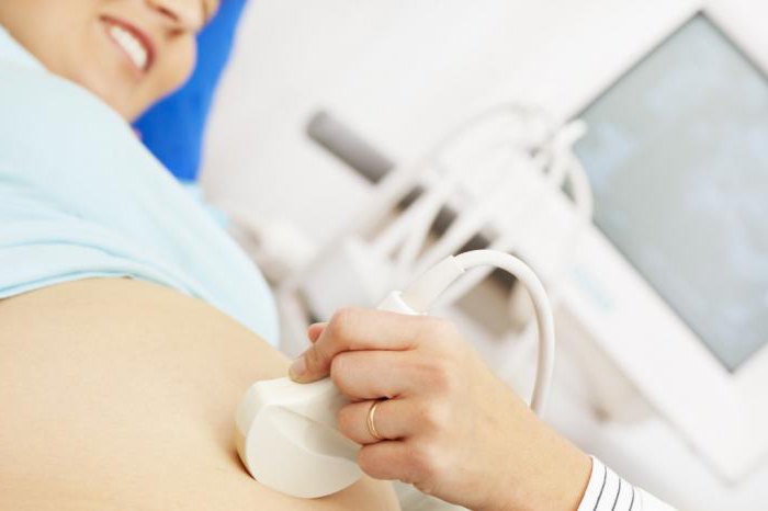
Pregnant Examination
The best option for a full examination of pregnant women is considered to be computer sonography. Such equipment allows you to carefully examine the condition of the fetal organs, starting with a four-centimeter size. The examination is completely safe. From the fourth week of pregnancy, it helps to listen to the heartbeat, to identify and eliminate the threat of miscarriage, to clarify the gestational age, to determine developmental delays and other abnormalities.
Patency of the fallopian tubes
The need to assess the patency of the fallopian tubes arises in women who cannot become pregnant for a long time. Sonography of the fallopian tubes is one of the methods for assessing the reasons why an egg cannot meet with a sperm.
First, the doctor examines the condition of the uterine cavity, makes sure that the woman is not pregnant and does not have adhesions, polyps and nodes. Then, physiological saline is injected into the cervical canal, and the patency of the fallopian tubes is assessed using sonography. If the tubes are passable, then the fluid drains from both sides of the organ into the abdominal cavity. If the fluid does not drain, but fills the segment of the fallopian tube and uterus, then the tube is impassable. Ultrasound allows you to accurately determine the location of the block.
If the patient understands the meaning of the term “sonography”, what it is and why the doctor ordered an examination, then he does not feel fear, is ready to fulfill the necessary requirements of the doctor and correctly refers to the procedure. Much depends on it. Because often, instead of examination and treatment, frightened patients turn to charlatans and “healers”, wasting precious time.