X-ray studies are firmly established in the practice of medical examination and diagnosis. The accessibility and informational content of these methods made them ubiquitous, and some even mandatory for preventive purposes. Fluorography is an examination that every citizen of our country must undergo once a year after reaching the age of 18 with the aim of preventing diseases, and it is it that causes the most complaints because of the fear of radiation. Is there any reason to be afraid of her? And how is fluorography different from lung x-rays?
What is x-ray radiation?
X-rays are a type of electromagnetic radiation with a wavelength of 0.005 to 10 nanometers. According to the characteristics, they somewhat coincide with gamma rays, but have a different origin. There are 2 types of radiation - soft and hard. The latter is used in medicine for diagnostic purposes.
Since it is impossible to focus the X-rays , during the examination, a radiating tube is directed at the patient and a receiving sensitive screen is placed behind him. An image will then be taken from it.
In clinics, fluorography is performed for prophylactic purposes. How is this examination different from x-rays? With the direct passage of rays, the structure of the organ is displayed on the screen, and with fluorography, its shadow is reflected, reflected from the fluorescent screen. Devices for these types of studies vary in design.
Fluorography determination
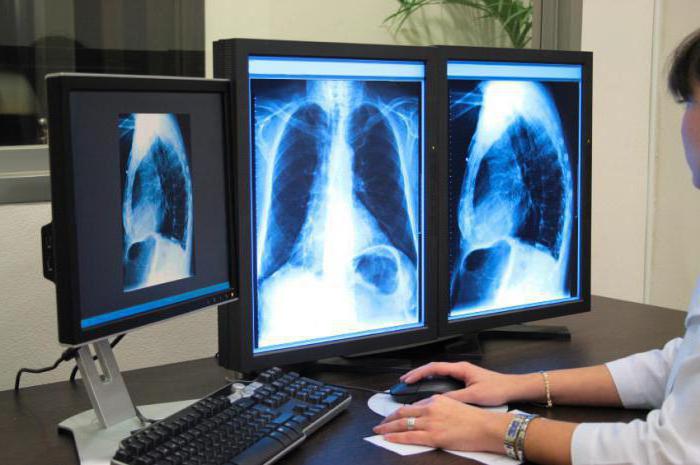
Fluorography is an X-ray examination of the chest organs, in which the image in the image is obtained by the reflected method. In the last decade, the digital version of the survey has become widespread, in which, instead of a picture, the result is displayed immediately on a computer screen, and then a description is made.
Indications for examination
This method is used for screening purposes, that is, if necessary, to examine a large number of the population in order to obtain results of a high degree of reliability in a short time. The identification of cases of tuberculosis is the main goal for which compulsory fluorography was once introduced. What distinguishes this examination technically from x-rays is its low resolution. However, it can be used to detect the presence of foreign bodies, fibrosis, developed inflammation, tumors, cavities, and the presence of infiltrates (seals).
Chest x-ray
Chest x-ray is a non-invasive method for examining tissues and organs using the same rays. The result is displayed on a film shot. This examination is also radiological. What is the difference between fluorography and chest x-ray for a simple layman, is the size of the finished result - instead of a small illegible square, a developed film of 35 x 35 cm is issued.
Indications for lung X-ray
Radiography as a more detailed examination is prescribed to identify inflammatory processes, anomalies of the anatomical structures, with suspected tumors of a different nature. Rarely is it used to see the location of the heart relative to other organs of the mediastinum.
What is the difference between fluorography and x-ray? The difference lies in the information content of the images and the detail of the resulting image. A classic x-ray makes it possible to see objects (seals, cavities, foreign bodies) up to 5 mm in diameter, while fluorography shows predominantly large changes. In complex diagnostic cases, only an extended examination will be used.
Radiation doses
Many are concerned about the health hazards of examinations. Patients are afraid that undergoing a routine or preventive examination may adversely affect their bodies. Of course, there is some harm from x-ray exposure, but not so serious.
The permissible
radiation dose per year without damage to health is 5 mSv (millisievert). In film radiography, a single dose is 0.1 mSv, which is 50 times less than the annual norm. Fluorography gives a slightly higher exposure. What distinguishes this examination from X-rays is the rigidity of the rays passing through the body, which is why a single dose increases to 0.5 mSv. Compared with permissible irradiation during the year, this is still not so much.
Digital technology to replace the film
The development of medical equipment has also affected the quality of X-ray equipment. Digital devices are being introduced everywhere in place of installations made in the last century that output the result only on tape. For patients, this innovation is good in that the radiation dose is significantly reduced. Digital exposure requires less exposure than film exposure. The well-known “hold your breath” during the examination is connected with the fact that when you inhale the soft tissues are displaced, “smearing” the shadows in the picture. But it is with the film result that fluorography is mainly done.
What is the difference from an X-ray made by the usual method, examination on a digital apparatus? First of all, by a decrease in radiation exposure. The effective equivalent dose obtained during digital fluorography is 0.05 mSv. A similar parameter with a chest x-ray will be 0.075 mSv (instead of the standard 0.15 mSv). Therefore, in order to maintain health it is more advisable to choose more modern examination methods.
Saving time is the second answer to the question of how fluorography differs from lung X-ray, made in the digital version. To obtain the result, you do not need to wait for the development of the image, so that later it can be described by a specialist.
Which method to choose?
Some people, having received a referral for a preventive annual examination, do not know what to choose - X-ray or fluorography of the lungs. If there are no complaints about the work of the respiratory system, then taking a large picture does not make much sense. If it is possible to do digital fluorography - do it, this will protect the body from an extra dose of radiation.
A doctor who suspects pneumonia or a serious disease of the mediastinal organs does not have the right to make a final diagnosis without confirmation by instrumental examination. In the presence of pathologies, questions about which is better - X-ray of the lungs or fluorography, therapists and pulmonologists are not asked. Every detail that research can give is important to them. Therefore, with a developed clinical picture of pneumonia, suspected tuberculosis or tumor process, the patient is sent for x-ray, more often in several projections.

If there is a prerequisite for the development of pulmonary diseases in the history, for example, the patient actively smokes or his work is associated with harm to the respiratory tract (welding, metal casting, chemical industry), the examination should be done regularly to prevent serious pathologies from developing. Twice a year, employees of tuberculosis dispensaries and hospitals are required to have x-ray or chest x-ray. What to choose, your doctor will tell you.
Contraindications for examination
Due to radiation effects on the body, X-ray examination of certain categories of patients should be carried out with caution or not done at all.
Individual organs react sharply to radiation, giving clinical pathology. Sex cells are especially sensitive, therefore it is not recommended to excessively irradiate the pelvic area. X-rays adversely affect the cells of the red bone marrow, disrupting their division and growth. The thyroid and thymus glands are also sensitive to all types of radiation, so when examining you need to keep your neck above the level of the radiating tube.
It is strongly not recommended to do an x-ray to pregnant women, as it affects the development of tissues and organs of the fetus. An exception is made only in case of a threat to the life of the expectant mother. Extensive X-ray studies are not recommended for children under 12 years of age, but it is allowed to take photographs of the limbs and maxillofacial region according to indications when using protective equipment.