The knee joint, the structure of which should be well known to every person involved in sports, is the largest in the human body. It is formed by three bones. The structure of a person’s knee joint is determined by its location. The ends of the bones that form its structure are covered with a very dense cartilage tissue up to 6 mm thick. This provides one of the main functions of the articulation - shock absorption when walking.
Knee joint, structure
The photo shows us the main structures of this joint: muscles, bones, menisci, ligaments (cruciform), nerves and blood vessels. We begin to consider its structure with bones. The joint is formed by three bones. Two long ones - tubular tibial and femoral. The third is the patella. It is rounded and quite small. Located in front. The femur below forms the condyles - protrusions covered with cartilage. These protrusions are in contact with the so-called tibial plateau, which, in turn, consists of two halves. The patella moves in a groove-shaped depression formed by the condyles. This notch is also called patellofemoral. The fibula is located on the side of the tibia. She does not participate in the formation of the knee joint.
The structure and importance of cartilage
The function of this fabric is to absorb shock load, reduce friction during movements. It is necessary where two bone surfaces rub against each other. The articular cartilage is very dense. In the knee joint, it covers not only the ends of the femur and tibia, but also the surface of the patella. Cartilage is of several types. In the knee joint - hyaline. A feature of this tissue is the high water content in the intercellular substance. This provides elasticity and helps protect the knee joint from damage.
The structure of ligaments and menisci
Dense connective tissue formations that fix the ends of the bones are called ligaments. In the case of the knee joint, its capsule is strengthened by two such structures from the outside - medial and lateral. And two inside - anterior and posterior cruciate. They limit excessive movement in the anteroposterior direction, preventing it from slipping relative to the femur. All ligaments of the knee are extremely important for its stable operation. Between the femur and the tibia are two more formations, called menisci. They can also be called cartilage, although their structure differs from the structure of hyaluronic, covering the articular surfaces. Menisci fill the space between the tibial plateau and the articular end of the femur.
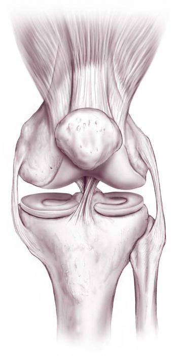
They seem to serve as an elastic pad, redistributing weight. Were it not for them, all its heaviness would have concentrated at one point on the tibial plateau. Two types of menisci (medial and lateral) are connected by a transverse ligament. Lateral (external) is less likely to be damaged due to its greater mobility. The inner (medial) meniscus is located near the inner lateral ligament and has less lability. This is due to its frequent trauma. In the center of the meniscus is thicker than at the edges - this forms a small cavity on the tibial plateau and makes the joint more stable. If there were no ligaments, we would have a much greater imbalance in the lower limb and more often would injure the knee joint. The structure of the supporting elements of the knee provides him stability
Synovial bags
They lie along the muscles and tendons. The largest is the patella (under the tendon of the quadriceps muscle), it hardly communicates with the joint cavity. A deep popliteal bag is located behind, in the thickness of the joint - several more smaller ones. When some of them are filled with intraarticular fluid, cysts may form.
Muscles involved in flexion and extension of the joint
The quadriceps is located on the front surface of the thigh. With its reduction, the leg in the knee joint is unbent. The patella lies in the thickness of the tendon, serving as a fulcrum and changing the direction of movement if necessary. It increases the strength of said muscle. The flexors of the lower leg (on the back of the thigh and near the knee) bend the leg in the knee joint.
Innervation
Consider the popliteal nerve. It is the largest of those located on the back of the joint. This nerve is a branch of the sciatic. It provides sensitive and motor innervation of the joint capsule. Above the joint, it is divided into the tibial and peroneal nerves. They are worth mentioning because with a knee injury they are often damaged. Also, the back of the capsule innervates the obturator nerve. Some branches of the tibial nerve provide sensitivity to its posterior part. The fibular innervates the posterior and anteroposterior surface. This is due to the fact that in the body there are few of the same mobile formations as the knee joint - the structure and innervation with a large number of overlapping zones provides high sensitivity.
Blood supply
The vast vascular network surrounding the knee consists of four large arteries that are interconnected and form the vascular plexus (there are about 13 such networks on the surface of the joint) and inside it. The first and largest artery is the femoral. The popliteal, deep and anterior tibial is slightly smaller. All of them develop collateral circulation in the event that one of the vessels is bandaged. The anatomical structure of the popliteal artery can be easily imagined by dividing it into three sections. The first is the topmost. Dressing is best done at the second level. Superficial veins in the knee joint are located in two layers. The deeper is represented by a large saphenous vein. Superficial - venous network from the incremental. The latter is not found in every person. A small saphenous vein departs from the posterior surface of the knee joint. Sometimes it goes with one trunk, and sometimes two. The place of its confluence also varies, but more often falls into the popliteal.