Due to the high activity, load, as well as other external and internal factors, the knee joints can be affected by a variety of pathological processes. The most common are the so-called degenerative-dystrophic conditions (gonarthrosis, both post-traumatic and idiopathic - that is, that arose for an unknown reason) and arthritis (rheumatic, infectious). Also, various cartilage lesions — chondropathies — are known to medicine due to both mechanical factors and a genetic predisposition.
Unfortunately, oncological processes in this anatomical area are also frequent. Some diseases, for example, bleeding disorders (primarily hemophilia) cause constant extensive bleeding into the joint cavity. This condition is called hemarthrosis. I must say that the knee joints are one of the most active joints in the musculoskeletal system of a person, together with the hips they carry the weight of the whole body every day. That is why gonarthrosis develops in individuals with obesity in the first place - cartilage is very quickly “worn out” and destroyed. On the other hand, the “activity” of the knee joints causes their frequent injuries. Among the most common injuries are tears of the ligaments and menisci.
Diagnosis of knee problems
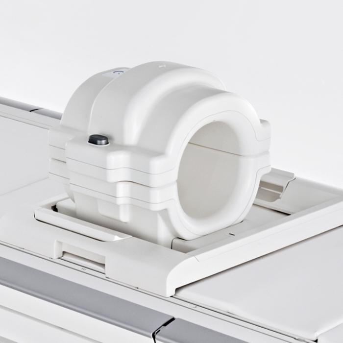
Of course, any treatment should be preceded by a diagnosis. One of the most effective methods for diagnosing injuries and diseases affecting the knee joints is MRI - magnetic resonance imaging. This is a common and safe modern medical technology, it is actively used in many clinics around the world. And its inventors P. Mansfield and P. Lauterbur received in 2003 the Nobel Prize. MRI of the knee allows you to visualize the smallest anatomical details, to detect early signs of pathological changes (especially in cartilage). Each element of the joint can be viewed at arbitrary magnification and in the required projection. The technology allows you to "remove" the patella (patella) during layering, gaining access to any internal structure.
How not to make a mistake?
Examination of the knee joints is necessary carefully and comprehensively. In American medicine, the special term “vomit” has even appeared (in Russian - vomiting). It is composed of the first letters of the phrase, which can be translated in this way: "the victim of medical imaging technology." This is the name of the doctors who make diagnostic errors, trusting tomograms and images too much and neglecting the clinical examination, talking with the patient, and an integrated systematic approach.
Despite the advantages and possibilities of MRI, the doctor must carefully question the patient, perform the so-called physical examination of the joint (that is, check various symptoms and signs of the disease), prescribe a general and biochemical blood and urine tests, and begin visualizing the joint with radiography. And only after all of the above, magnetic resonance imaging is prescribed.
Perhaps the most important advantage of MRI is that it allows you to get clear images of the so-called soft tissue structures - menisci and ligaments of the knee joint. It is these images, coupled with the above studies that allow the doctor to make the correct correct diagnosis and prescribe the optimal treatment.
In conclusion, we say that the only contraindication to MRI is the presence of implants and pacemakers in the body, as well as the first trimester of pregnancy. The patient is not exposed to radiation during the study, and it lasts only 30-40 minutes. The technological feature of the MRI device is a loud sharp noise. It's not worth being scared. We also add that there are no complications during and after this procedure.