The nervous system consists not only of neurons and their processes. 40% of it is represented by glial cells, which play an important role in its life. They literally limit the brain and nervous system from the rest of the body’s environments and ensure its autonomous functioning, which is really important for humans and other animals that have a central nervous system. Moreover, neuroglia cells are able to divide, which distinguishes them from neurons.
The general concept of neuroglia
The collection of glial cells is called neuroglia. These are special cell populations that are located in the central nervous system and on the periphery. They maintain the shape of the brain and spinal cord, as well as supply it with nutrients. It is known that in the central nervous system, due to the presence of the blood-brain barrier, there are no immune reactions. However, when a foreign antigen enters the brain or spinal cord, as well as into the cerebrospinal fluid, the glial cell, a reduced analogue of the macrophage of peripheral tissues, phagocytes it. Moreover, it is the separation of the brain from peripheral tissues that provides neuroglia.
Brain immunity
The brain, where many biochemical reactions take place, which means that a mass of immunogenic substances are formed, must be protected from humoral immunity. It is important to understand that brain neuronal tissue is very sensitive to damage, after which neurons are only partially restored. This means that the appearance of a place in the central nervous system where the local immune reaction will take place will also result in the death of some surrounding cells or demyelination of the processes of neurons.
At the periphery of the body, this damage to somatic cells will soon be filled with newly formed ones. And in the brain it is impossible to restore the function of a lost neuron. And it is neuroglia that limits the brain from contact with the immune system, for which the central nervous system is a huge number of foreign antigens.
Glial cell classification
Glial cells are divided into two types depending on morphology and origin. Microglia and macroglia cells are isolated. The first type of cells originates from the mesoderm leaf. These are small cells with numerous processes that can phagocytize solids. Macroglia is a derivative of ectoderm. The glial cell of macroglia is divided into several species depending on morphology. Ependymal and astrocytic cells, as well as oligodendrocytes, are isolated. Such types of cell populations are also divided into several types.
Ependymal glial cell
Ependymal glial cells are found in specific areas of the central nervous system. They form the endothelial lining of the cerebral ventricles and the central spinal canal. They take their origin in embryogenesis from the ectoderm, and therefore represent a special type of neuroepithelium. It is multilayer and performs a number of functions:
- supporting: makes up the mechanical frame of the ventricles, which is also supported by the hydrostatic pressure of the cerebrospinal fluid;
- secretory: secretes some chemicals into the cerebrospinal fluid;
- demarcation: separates the medulla from the cerebrospinal fluid.
Types of Ependymocytes
Among ependymocytes, there are also their own species. These are ependymocytes of the 1st and 2nd order, as well as tanicites. The first form the initial (basal) layer of the ependymal membrane, and ependymocytes lie in the second layer above them. It is important that the 1st order ependymal glial cell is involved in the formation of the hematoglyphic barrier (between the blood and the internal environment of the ventricles). 2nd order ependymocytes have villi oriented in the direction of cerebrospinal fluid flow. There are also tanicites, which are receptor cells.
They are located in the lateral areas of the bottom of the 3rd cerebral ventricle. Having microvilli on the apical side and one process on the basal, they can transmit information to neurons about the composition of the cerebrospinal fluid. In this case, the cerebrospinal fluid itself, through small numerous slit-like openings between the 1st and 2nd order ependymocytes, can directly reach the neurons. This suggests that ependyma is a special type of epithelium. Its functional, but not morphological analogue on the periphery of the body is the endothelium of blood vessels.
Oligodendrocytes
Oligodendrocytes are the types of glial cells that surround a neuron and its processes. They are found both in the central nervous system and near peripheral mixed and autonomic nerves. Oligodendrocytes themselves are polygonal cells equipped with 1-5 processes. They interlock with each other, isolating the neuron from the internal environment of the body and providing conditions for the nervous conduct and generation of impulses. There are three types of oligodendrocytes, which differ in morphology:
- a central cell located near the body of the brain neuron;
- a satellite cell surrounding the body of a neuron in the peripheral ganglion;
- Schwann cell, covering the neuronal process and forming its myelin sheath.
Oligodendrocytic glial cells are found both in the brain and spinal cord, and in peripheral nerves. Moreover, it is still unknown how the satellite cell differs from the central one. Given that the genetic material is the same for all cells of the body, except for sex, then probably these oligodendrocytes can mutually replace each other. The functions of oligodendrocytes are as follows:
- supporting;
- insulating;
- separation;
- trophic.
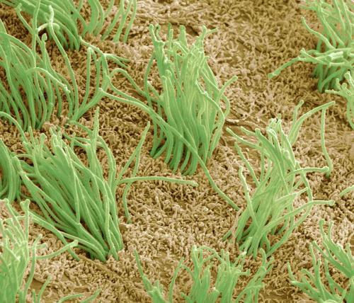
Astrocytes
Astrocytes are the glial cells of the brain that make up the brain matter. They are star-shaped and small in size, although they are larger than microglia cells. However, there are only two types of astrocytes: fibrous and protoplasmic. The first type of cells is located in the white and gray matter of the brain, although they are much more in white.
This means that they are most common in those areas where there is a significant number of neuronal myelinated processes. Protoplasmic astrocytes are also glial cells: they are found in the white and gray matter of the brain, but they are much more in gray. Therefore, their function is to create support for the bodies of neurons and the structural organization of the blood-brain barrier.
Microglia
Microglial cells are the last type of neuroglia. However, unlike all other cells of the central nervous system, they are of mesodermal origin and are special types of monocytes. Their precursors are stem blood cells. Due to the structural features of neurons and their processes, glial cells are responsible for the immune responses in the central nervous system. And their functions are almost similar to those of tissue macrophages. They are responsible for phagocytosis and recognition and presentation of antigen.
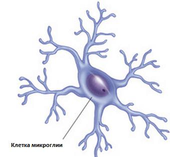
Microglia contains special types of glial cells that have differentiation cluster receptors, which confirms their bone marrow origin and the implementation of immune functions in the central nervous system. They are also responsible for the development of demyelinating diseases, Alzheimer's disease and Parkinson's syndrome. However, the cell itself is only a way to implement the pathological process. Therefore, it is likely that when it is possible to find the mechanism of microglia activation, the development of these diseases will be suppressed.