The skeleton, a photo of which will be presented below, is a collection of bone elements of the body. The word itself has ancient Greek roots. In translation, the term means "dried." The skeleton is considered a passive part of the musculoskeletal system. It develops from mesenchyme. Next, we consider in more detail the skeleton: structure, functions, etc.
Gender Features
Before talking about what functions the skeleton performs, a number of distinguishing features of this part of the body should be noted. In particular, some sexual features of the structure are of interest. In total, there are 206 bones that make up the skeleton (the photo illustrates all its elements). Almost all are connected into a single whole through joints, ligaments and other joints. The structure of the skeleton of men and women is generally the same. There are no cardinal differences between them. However, differences are found only in a few altered forms or sizes of the individual elements and systems that they make up. The most obvious differences that the structure of the skeleton of men and women have, for example, are that the bones of the fingers and limbs in the former are somewhat longer and thicker than in the latter. In this case, tuberosity (areas of fixation of muscle fibers) are expressed, as a rule, stronger in men. In women, the pelvis is wider, and the chest is narrower. As for gender differences in the skull, they are also insignificant. In this regard, it is often difficult for specialists to determine to whom it belongs: a woman or a man. At the same time, the eyebrows and tuberosity protrude more strongly in the latter, the eye sockets are larger, the paranasal sinuses are more pronounced. In the male skull, the bone elements are somewhat thicker than in the female. The anteroposterior (longitudinal) and vertical parameters of this part of the skeleton in men are greater. The capacity of the skull of women is about 1300 cm 3 . In men, this indicator is also greater - 1450 cm 3 . This difference is due to the smaller overall size of the female body.
Head department
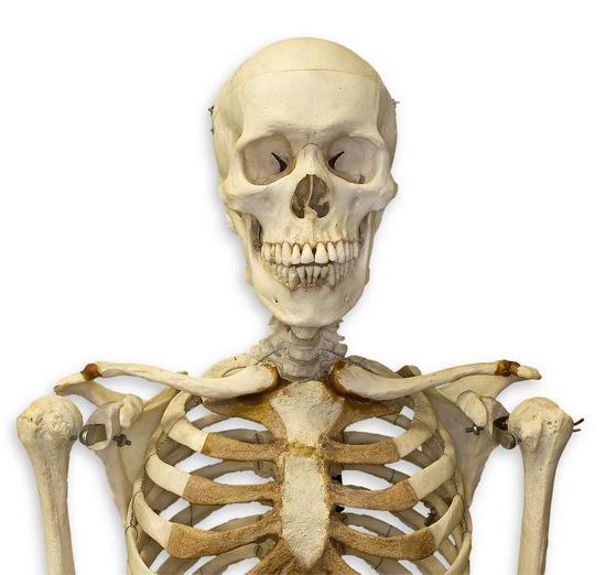
Two zones are distinguished in the skeleton. In particular, the trunk and head sections are present in it. The latter, in turn, includes the facial and brain parts. The brain part contains 2 temporal, 2 parietal, frontal, occipital and partially ethmoid bones. The composition of the facial section contains the upper jaw (paired) and lower. In their holes teeth are fixed.
Spine
In this section, coccygeal (4-5 pcs.), Sacral (5), lumbar (5), thoracic (12) and cervical (7) segments are distinguished. Vertebral arches form the spinal canal. The pillar itself has four bends. Thanks to this, it is possible to carry out an indirect function of the skeleton associated with upright posture. Between the vertebrae are elastic plates. They contribute to improving the flexibility of the spine. The appearance of bends of the pillar is caused by the need to mitigate tremors during movement: running, walking, jumping. Due to this, the spinal cord and internal organs are not subjected to concussion. A channel runs inside the spine. It surrounds the spinal cord.
Rib cage
It includes the sternum, 12 segments of the second spine, as well as 12 costal pairs. The first 10 of them are connected to the sternum by cartilage, the last two do not have joints with it. Thanks to the chest, the protective function of the skeleton is possible. In particular, it ensures the preservation of the heart and organs of the bronchopulmonary and partially digestive systems. The costal plates on the back have a movable joint with the vertebrae, while the front (except for the lower two pairs) are connected to the sternum by means of flexible cartilage. Due to this, the chest can narrow or expand when breathing.
Upper limbs
In this part, the humerus, forearm (ulnar and radial elements), wrist, five metacarpal segments and digital phalanges are present. In general, three departments are distinguished in the skeleton of the arm . These include the hand, forearm and shoulder. The latter is formed by a long bone. The hand is connected to the forearm and consists of small carpal elements, the metacarpus forming the palm, and also flexible flexible fingers. The attachment of the upper limbs to the body is carried out by means of clavicles and shoulder blades. They form the shoulder girdle.
Lower limbs
2 pelvic bones are secreted in this part of the skeleton. Each of them includes sciatic, pubic and iliac elements fused with each other. Also, the hip is referred to the lower extremity belt. It is formed by the corresponding bone of the same name. This element is considered the largest of all in the skeleton. The leg is also secreted in the leg. The structure of this department includes two tibia - large and small. Hangs the lower limb of the foot. It consists of several bones, the largest of which is the heel. The articulation with the body is carried out through the pelvic elements. In humans, these bones are more massive and wider than in animals. The joints act as connecting elements of the limbs.
Joint Types
There are only three of them. In the skeleton, the bones can be connected movably, semi-mobile or motionless. Jointing according to the latter type is characteristic of cranial elements (except for the lower jaw). The ribs with the sternum and vertebrae are semi-mobile. Ligaments and cartilage act as articulation elements. A movable joint is characteristic of joints. Each of them has a surface, a fluid present in the cavity, and a bag. As a rule, joints are strengthened by ligaments. Due to them, the amplitude of motion is limited. Joint fluid reduces friction of bone elements during movement.
What functions does the skeleton perform?
This part of the body has two tasks: biological and mechanical. In connection with the solution of the latter problem, the following functions of the human skeleton are distinguished:
- Propulsion. This task is carried out indirectly, since the elements of the skeleton are used to attach muscle fibers.
- The supporting function of the skeleton. Bone elements and their joints make up the skeleton. Attached to it are organs and soft tissues.
- Spring. Due to the presence of articular cartilage and a number of structural features (bends of the spine, arch of the foot), depreciation is carried out. As a result, tremors are eliminated and tremors are mitigated.
- Protective. Bone formations are present in the skeleton, due to which the preservation of important organs is ensured. In particular, the skull protects the brain, the sternum - the heart, lungs and some other organs, the spine - the spinal structure.
Biological functions of the human skeleton:
- Hematopoietic. Bone marrow is located in the bones. It acts as a source of blood cells.
- Reserving. Bone elements serve as a depot for a large number of inorganic substances. These, in particular, include iron, magnesium, calcium, phosphorus. In this regard, the bones are involved in maintaining a stable mineral composition within the body.
Damage
In case of improper body position for a long period (for example, prolonged sitting with your head bowed at the table, uncomfortable posture, etc.), as well as against the background of a number of hereditary reasons (especially in combination with errors in nutrition, insufficient physical development), a violation may occur retention function of the skeleton. In the early stages, this phenomenon can be eliminated quickly enough. Nevertheless, it is better to prevent it. To do this, experts recommend choosing a comfortable position at work, regularly engage in sports, gymnastics, swimming and other forms.
Another fairly common pathological condition is deformity of the foot. Against the background of this phenomenon, there is a violation of the motor function of the skeleton. Deformation of the foot can occur under the influence of diseases, be the result of injuries or prolonged overload of the foot in the process of growth of the body.
Under the influence of strong physical activity, a bone fracture can occur. This type of injury can be closed or open (with a wound). About 3/4 of all fractures occur in the arms and legs. The main sign of injury is severe pain. A fracture can provoke subsequent bone deformation, impaired function of the department in which it is located. If there is a suspicion of a fracture, the victim must be provided with an ambulance and hospitalized. Before taking any action, the patient is sent for x-ray examination. During the diagnosis, a fracture localization site, the presence and displacement of bone fragments are revealed.