Ultrasound diagnostics and magnetic resonance imaging are quite popular research methods today. However, a large number of people do not quite understand why both methods are intended and what is their difference. Which is better - ultrasound or MRI and in what cases?
Ultrasound diagnostics
Ultrasound is carried out using a special apparatus, which through ultrasonic waves displays information from the sensor to the screen. This diagnostic method is considered safe, since exposure is minimal. The benefits of ultrasound:
- the lack of strong exposure makes it possible to diagnose even pregnant women and newborns;
- there is no quantitative limit of procedures, therefore, it is possible to carry out an ultrasound without harm to health as many times as the correct diagnosis requires;
- accessibility - there is an ultrasound machine in almost every, even a small hospital;
- low cost;
- high research efficiency.
Ultrasound or MRI of the pelvis - which is better? It is more convenient to examine the peritoneal organs with an ultrasound device, since the device is effective for diagnosing internal organs. Unfortunately, ultrasound is of little use for examining the bone structure.
Magnetic resonance imaging
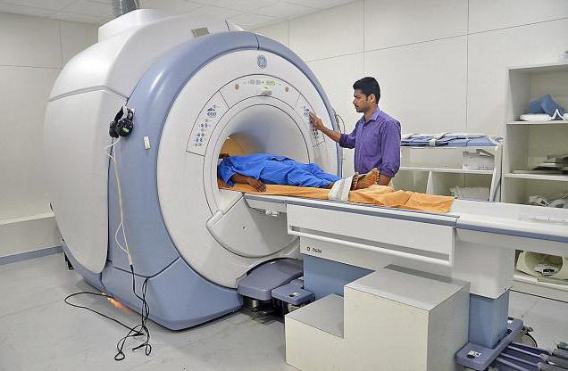
There is no single answer to the question of which is better - ultrasound or MRI. However, each of the methods has its own characteristics. MRI is a modern method of hardware diagnostics, which is appreciated for its high information content. The device creates magnetic waves, with the help of which the necessary information is displayed on the screen. The study takes from 20 to 50 minutes, depending on the object. At this time, the patient should lie still so that the information is most accurate. Thanks to the MRI device, there is a possibility of timely detection of abnormalities and their treatment.
Most often, using magnetic resonance imaging, the pathologies of the following systems are examined:
- cardiovascular;
- musculoskeletal system;
- cerebral vessels.
The study has the following features:
- Security. In this case, the patient receives a minimum dose of radiation, which does not affect well-being and health.
- High information content and detail of the information received.
- The ability to identify hidden pathologies that were not detected using other diagnostic methods.
- The possibility of repeated use in a short period of time.
However, a study cannot be prescribed in such cases:
- the presence of metal implants or a pacemaker, which may be affected by a magnetic field;
- heart or kidney failure;
- first trimester of pregnancy;
- lactation period;
- allergic reactions to a contrast drug, if there is a need for the introduction of contrast.
In addition, tomography is not recommended for patients with claustrophobia, as well as tattoos that contain metal particles in their dyes. Among the disadvantages of the method, a rather high cost of the procedure can be noted.
Differences of ultrasound from MRI
Without the comparative characteristics of the two diagnostic methods, it is impossible to determine which is better - ultrasound or MRI. There are three main differences:
- The sensitivity of the ultrasound machine is less than that of the tomograph, so it is most often used to monitor the course of the disease. With its help, large tumors can be detected.
- Magnetic tomography is highly accurate information. With its help, even small formations can be detected at an early stage of their development.
- Not only the functionality and accuracy of diagnostics is radically different, but also their cost. Tomography is an expensive procedure that not everyone can afford, so most often people are forced to use ultrasound.
Don't know which is better - an ultrasound or an MRI? First of all, pay attention to the scope. Thus, ultrasound is more informative for the study of soft tissues, and MRI for bone and vascular systems.
The accuracy of diagnostic methods
As already noted, magnetic resonance imaging is highly accurate, therefore, it is recommended to use it for the diagnosis of diseases of the skeletal structure and blood vessels. However, the patient does not always know what is best - ultrasound or MRI. Doctors definitely advise to do an MRI.
Regarding the cardiovascular system, MRI is fully capable of giving the most detailed information about the smallest changes in the vessels, as well as detecting pathology at the initial stage of its development. Which is better - ultrasound of vessels or MRI of vessels? Tomography in this case will be preferable, since it will provide the most accurate information. For example, if it is necessary to study the vessels of the brain, a tomography is performed with contrast, which will allow you to better see the outlines of the blood flow. The drug for contrasting is produced on the basis of substances that are completely safe for humans.

Which is better - ultrasound or MRI of the head? It all depends on what part of the head should be examined. If we are talking about vessels, then it is recommended that both MRI and ultrasound, but the bone structure can be examined only with the help of tomography. Among ultrasound diagnostic techniques, Dopplerography can also be found, which can also help assess the condition of the vessels. Indications for her appointment are as follows:
- frequent dizziness;
- migraine;
- loss of consciousness;
- sensation of tinnitus;
- stroke;
- sharp visual impairment;
- hypertension;
- disorders in the field of neurology.
It is important to determine in a timely manner what is best done - ultrasound or MRI. In each case, the doctor makes the decision to make the correct diagnosis as soon as possible.
The choice of method for the diagnosis of the spine
Most often, injuries lead to diseases of the spine. How to act in this case? Which is better - ultrasound or MRI? It is definitely necessary to do a tomography, since ultrasound is not sufficiently informative when it comes to bone structure. In addition, MRI helps to identify the formation of tumors, arthrosis, and inflammatory processes.
Ultrasound diagnostics for diseases of the spine is prescribed in the following cases:
- numbness of the limbs;
- severe pain in the area of internal organs;
- high or low blood pressure.
Ultrasound is used to study the cervical, thoracic, lumbar spine, as well as the connective tissue of the knee and elbow joints.
Examination of the pelvic organs
Ultrasound is a popular diagnostic method for examining the pelvic organs. Ultrasound rays pass well through the soft tissues and body fluids, so the information content of such a study will be at a high level. In this case, a sufficiently vast space can be examined with a sensor. MRI of the pelvic organs is prescribed in the following cases:
- The occurrence of inflammatory processes.
- Suspicion of benign or malignant tumors.
- Mechanical damage to bone tissue.
- A variety of vascular pathologies.
If we are talking about the pelvic organs, then tomography is assigned equally to both men and women. The following organs are examined:
- bladder;
- uterus;
- ovaries;
- prostate;
- seminal vesicle and ducts.
MRI provides more detailed information about the state of organs.
Examination of the peritoneal organs
One of the organs often suffering from diseases is the liver. That is why many people think about what is better - ultrasound or MRI of the liver? Often the answer to this question can be given by the attending doctor, who directs to research, based on the symptoms of the disease. In addition, both ultrasound and MRI are prescribed for various pathologies of the gastrointestinal tract, heart problems, and kidney diseases.
Finally
There is no definite answer about what is best from the two diagnostics. Each direction is carried out taking into account the capabilities of the hospital, patient, as well as medical indications. Both methods are good enough, but have minor flaws.