In the human body there are several types of different tissues. All of them play a role in our life. One of the most important is connective tissue. Its specific gravity is about 50% of the mass of a person. It is a connecting link connecting all the tissues of our body. Many functions of the human body depend on its condition. Various types of connective tissue are discussed below.
General information
Connective tissue, the structure and functions of which have been studied for many centuries, is responsible for the work of many organs and their systems. Its specific gravity is from 60 to 90% of their mass. It forms the supporting frame, called the stroma, and the outer integument of the organs, called the dermis. The main features of connective tissue:
- common origin from mesenchyme;
- structural similarity;
- performance of supporting functions.
The main part of the solid connective tissue belongs to the fibrous type. It consists of elastin and collagen fibers. Together with the epithelium, connective tissue is an integral part of the skin. At the same time, it combines it with muscle fibers.
Connective tissue is strikingly different from others in that it is represented in the body by 4 different conditions:
- fibrous (ligaments, tendons, fascia);
- solid (bones);
- gel-like (cartilage, joints);
- liquid (lymph, blood; intercellular, synovial, cerebrospinal fluid).
Also representatives of this type of tissue are: sarcolemma, fat, extracellular matrix, iris, sclera, microglia.
The structure of connective tissue
It includes motionless cells (fibrocytes, fibroblasts) that make up the main substance. It also has fibrous masses. They are an intercellular substance. In addition, various free cells (fat, vagus, obese, etc.) are present in it. Connective tissue incorporates an extracellular matrix (base). The jelly-like consistency of this substance is due to its composition. The matrix is a highly hydrated gel formed by macromolecular compounds. They make up about 30% of the weight of the intercellular substance. At the same time, the remaining 70% is water.
Classification of connective tissue
The classification of this type of tissue is complicated by their variety. So, its main types are subdivided, in turn, into several separate groups. There are such types:
- Actually connective tissue, from which fibrous and specific are distinguished, characterized by special properties. The first is divided into: loose and dense (unformed and shaped), and the second - into fatty, reticular, mucous, pigmented.
- Skeletal, which is divided into cartilage and bone.
- Trophic, which includes blood and lymph.
Any connective tissue determines the functional and morphological integrity of the body. She has such characteristic features:
- tissue specialization;
- universality;
- multifunctionality;
- ability to adapt;
- polymorphism and multicomponent.
Common connective tissue functions
Different types of connective tissue perform the following functions:
- structural;
- ensuring water-salt balance;
- trophic;
- mechanical protection of the bones of the skull;
- formative (for example, the shape of the eyes is determined by the sclera);
- ensuring constancy of tissue permeability;
- mechanical support (cartilage and bone tissue, aponeurosis and tendon);
- protective (immunology and phagocytosis);
- plastic (adaptation to new environmental conditions, wound healing);
- homeostatic (participation in this important process of the body).
In a general sense, connective tissue functions:
- giving the human body shape, stability, strength;
- protection, coating and interconnection of internal organs.
The main function of the intercellular substance contained in the connective tissue is supporting. Its base provides a normal metabolism. Nerve and connective tissue provides the interaction of organs and various body systems, as well as their regulation.
The structure of various types of fabrics
The structure of connective tissue varies depending on its type. It consists of different cells and intercellular substance. A distinctive feature of such tissue is its high regenerative ability. It is characterized by ductility and good adaptation to changing environmental conditions. Any kind of connective tissue grows and develops due to the reproduction and transformation of young poorly differentiated cells. They come from mesenchyme, which is an embryonic tissue formed from the mesoderm (middle germinal leaf).
The intercellular substance, called the extracellular matrix, contains many different compounds (inorganic and organic). The consistency of connective tissue depends on their composition and quantity. Such substances as blood and lymph, in their composition contain intercellular substance in liquid form, called plasma. The cartilage matrix has the appearance of a gel. The intercellular substance of bones and tendon fibers are solid insoluble substances.
The intercellular matrix is represented by proteins such as elastin and collagen, glycoproteins and proteoglycans, glycosaminoglycans (GAG). It may include structural proteins laminin and fibronectin.
Loose and dense connective tissue
These types of connective tissue contain cells and intercellular matrix. In loose they are much more than in dense. In the latter, various fibers predominate. The functions of these tissues are determined by the ratio of cells and intercellular substance. Loose connective tissue performs primarily trophic function. Moreover, she also participates in supporting-mechanical activity. Cartilage, bone and dense fibrous connective tissue in the body perform a supporting-mechanical function. The rest are trophic and protective.
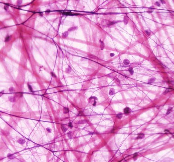
Loose fibrous connective tissue
Loose, unformed fibrous connective tissue, the structure and functions of which are determined by its cells, is found in all organs. In many of them, it forms the basis (stroma). It consists of collagen and elastic fibers, fibroplasts, macrophages, and a plasma cell. This tissue accompanies the blood vessels of the circulatory system. Through its loose fibers, there is a process of blood metabolism with cells, during which there is a transition of nutrients from it into tissues.
There are 3 kinds of fibers in the intercellular substance:
- Collagen, which go in different directions. These fibers have the form of straight and wave-like strands (constrictions). Their thickness is 1-4 microns.
- Elastic, which is slightly thicker than collagen fibers. They connect (anastomose) with each other, forming a broad-net network.
- Reticular, distinguished by their subtlety. They are intertwined in a mesh.
The cellular elements of loose fibrous tissue are:
- Fibroplasts, which are the most numerous. They have a fusiform shape. Many of them are equipped with processes. Fibroplasts are able to multiply. They take part in the formation of the main substance of this type of tissue, being the basis of its fibers. These cells produce elastin and collagen, as well as other substances related to the extracellular matrix. Inactive fibroplasts are called fibrocytes. Fibroclasts are cells that can digest and absorb the intercellular matrix. They are mature fibroplasts.
- Macrophages, which can be round, elongated and irregular in shape. These cells can absorb and digest pathogens and dead tissues, neutralize toxins. They are directly involved in the formation of immunity. They are divided into histocytes (in a calm state) and free (wandering) cells. Macrophages are distinguished by their ability to amoebiform movements. By their origin, they belong to blood monocytes.
- Fat cells that can accumulate a reserve in the form of drops in the cytoplasm. They have a spherical shape and are able to displace other structural units of tissues. In this case, dense fatty connective tissue is formed. It protects the body from heat loss. In humans, adipose tissue is mainly located under the skin, between the internal organs, in the omentum. It is divided into white and brown.
- Plasma cells located in the intestinal tissues, bone marrow and lymph nodes. These small structural units are distinguished by their round or oval shape. They play an important role in the activity of the body's defense systems. For example, in the synthesis of antibodies. Plasma cells produce blood globulins, which play an important role in the normal functioning of the body.
- Mast cells, often called tissue basophils, are characterized by their granularity. Their cytoplasm contains special granules. They come in a variety of shapes. Such cells are located in the tissues of all organs having a layer of unformed loose connective tissue. They include substances such as heparin, hyaluronic acid, histamine. Their direct purpose is the secretion of these substances and the regulation of microcirculation in tissues. They are considered immune cells of this type of tissue and respond to any inflammation and allergic reactions. Tissue basophils are concentrated around blood vessels and lymph nodes, under the skin, in bone marrow, spleen.
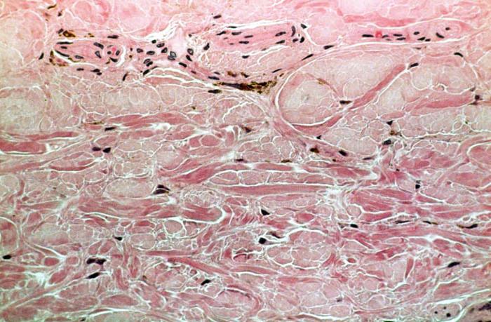
- Pigment cells (melanocytes) having a highly branched shape. They contain melanin. These cells are found in the skin and the iris of the eye. By origin, ectodermal cells are isolated, as well as derivatives of the so-called neural crest.
- Adveptive cells located along the blood vessels (capillaries). They are distinguished by their elongated shape and have a core in the center. These structural units can multiply and transform into other forms. It is at their expense that replenishment of dead cells of this tissue occurs.
Dense fibrous connective tissue
Connective tissue includes tissue:
- Dense unformed, which consists of a significant number of densely spaced fibers. It also includes a small number of cells located between them.
- Dense decorated, characterized by a special arrangement of connective tissue fibers. It is the main building material of ligaments and other formations in the body. So, for example, tendons are formed by densely arranged parallel bundles of collagen fibers, the spaces between which are filled with the main substance and a thin elastic network. Dense fibrous connective tissue of this type contains only fibrocyte cells.
Elastic fibrous is also isolated from it, from which some ligaments (vocal) are composed. The shells of round vessels, the walls of the trachea and bronchi are formed from them. In them, flattened or thick rounded elastic fibers are directed in parallel, while many of them have branches. The space between them is occupied by loose unformed connective tissue.
Cartilage
Connective cartilage tissue is formed by cells and a large volume of intercellular substance. It is designed to perform a mechanical function. There are 2 types of cells that make up this tissue:
- Chondrocytes having an oval shape and a nucleus. They are in capsules around which the intercellular substance is distributed.
- Chondroblasts, which are flattened young cells. They are located on the periphery of the cartilage.
Specialists divide the cartilage tissue into 3 types:
- Hyaline, found in various organs, such as ribs, joints, airways. The intercellular substance of such cartilage is translucent. It has a uniform consistency. Hyaline cartilage is covered with perichondrium. It has a bluish-white tint. It consists of the skeleton of the embryo.
- Elastic, which is the building material of the larynx, epiglottis, walls of the external auditory canals, the cartilaginous part of the auricle, and small bronchi. In its intercellular substance are developed elastic fibers. There is no calcium in this cartilage.
- Collagen, which is the basis of intervertebral discs, menisci, pubic articulation, sternoclavicular and mandibular joints. Its extracellular matrix includes a dense fibrous connective tissue, consisting of parallel bundles of collagen fibers.
This type of connective tissue, regardless of location in the body, has the same coverage. It is called the perichondrium. It consists of a dense fibrous tissue, which includes elastic and collagen fibers. It has a large number of nerves and blood vessels. Cartilage grows due to the transformation of the structural elements of the perichondrium. However, they are able to quickly transform. These structural elements turn into cartilage cells. This fabric has its own characteristics. So, the extracellular matrix of mature cartilage does not have blood vessels, so its nutrition is carried out by means of diffusion of substances from the perichondrium. This fabric is distinguished by its flexibility, it is resistant to pressure and has sufficient softness.
Bone connective tissue
Connective bone tissue is particularly hard. This is due to calcification of its intercellular substance. The main function of connective bone tissue is supporting-mechanical. All skeleton bones are built from it. The main structural elements of the fabric:
- Osteocytes (bone cells), which have a complex process shape. They have a compact core of a dark shade. These cells are located in bone cavities that follow the contours of osteocytes. Intercellular substance is located between them. These cells are unable to reproduce.
- Osteoblasts, which are a structural element of the bone. They have a rounded shape. Some of them have several cores. Osteoblasts are located in the periosteum.
- Osteoclasts, which are large multinucleated cells involved in the destruction of calcified bone and cartilage. Throughout human life, a change in the structure of this tissue occurs. At the same time, along with the process of decay, the formation of new elements occurs at the site of destruction and in the periosteum. Osteoclasts and osteoblasts are involved in this complex cell replacement.
Bone tissue contains an intercellular substance consisting of a basic amorphous substance. It contains ossein fibers that are not found in other organs. Connective tissue includes tissue:
- coarse fibrous, present in embryos;
- lamellar, available in children and adults.
This type of tissue consists of such a structural unit as a bone plate. It is formed by cells located in special capsules. Between them there is a fine fiber intercellular substance, which contains calcium salts. Ossein fibers having a significant thickness in the bone plates are arranged parallel to each other. They lie in a certain direction. Moreover, in adjacent bone plates, the fibers have a direction perpendicular to other elements. Thanks to this, a greater strength of this fabric is provided.
Bone plates located in different parts of the body are arranged in a certain order. They are the building material of all flat, tubular and mixed bones. In each of them, the plates are the basis of complex systems. For example, a tubular bone consists of 3 layers:
- An outer one in which the plates on the surface overlap with the next layer of these structural units. However, they do not form complete rings.
- The middle is formed by osteons, in which bone plates are formed around blood vessels. At the same time they are located concentrically.
- The inner, in which the layer of bone plates limits the space where the bone marrow is located.
Bones grow and regenerate due to the periosteum covering their outer surface, consisting of connective fine-fibrous tissue and osteoblasts. Mineral salts determine their strength. With a lack of vitamins or hormonal disorders, the calcium content is significantly reduced. The bones form the skeleton. Together with the joints, they represent the musculoskeletal system.
Diseases caused by connective tissue weakness
Insufficient strength of collagen fibers, weakness of the ligamentous apparatus can cause serious diseases such as scoliosis, flat feet, hypermobility of the joints, omission of organs, retinal detachment, blood diseases, sepsis, osteoporosis, osteochondrosis, gangrene, edema, rheumatism, cellulitis. Many experts attribute the weakening of immunity to the pathological condition of connective tissue, since the circulatory and lymphatic systems are responsible for it.