Among the main systems that make up the human body, the circulatory system occupies a special place. The structure of the circulatory system until the 16th century remained a mystery to scientists. Such outstanding thinkers as Aristotle, Galen, Harvey and many others worked on its solution. All their discoveries are summarized in a harmonious system of anatomical and physiological representations.
History reference
The Spanish scientist Servet and the English naturalist William Harvey played a special role in forming the correct ideas about what organs the human circulatory system consists of. The first managed to prove that blood from the right ventricle can enter the left atrium only through the network of blood vessels in the lungs. Harvey opened the so-called large circle (closed) circulation. Thus, a point was put in the question of whether the blood moves strictly in a closed system or not. The circulatory system of humans and mammals is closed.
It is also necessary to recall the works of the Italian doctor Malpigi, who discovered capillary circulation. Thanks to his research, it became clear how arterial blood turns into venous blood and vice versa. How does anatomy view this issue? The human circulatory system is a combination of such organs as the heart, blood vessels and auxiliary organs - red bone marrow, spleen and liver.
The heart is the main organ of the human circulatory system
Since ancient times, in all cultures without exception, the heart has been given a central role not only as an organ of the physical organism, but also as a spiritual reservoir of a person’s personality. In the expressions “heart friend”, “from the bottom of my heart”, “sadness at heart”, people showed the role of this organ in the formation of emotions and feelings.
But back to the anatomy and physiology of the heart. It is a muscular hollow organ, which is divided by an impenetrable septum into the left (cuboid, having an irregular shape) and right (regular shape) parts. It was the work of Servet and Harvey refuted the idea of Galen that there are special openings in the cardiac septum through which fluid connective tissue moves from one half to the other.
In fact, the presence of holes in the cardiac septum is a severe developmental pathology that relates to heart defects. It is diagnosed in the first days of a baby’s life, since when listening to a phonendoscope in the chest area, so-called noises (extraneous sounds arising from the mixing of arterial and venous blood) are clearly heard. This is a serious circulatory disorder, and only recently has it been amenable to operable treatment.
Heart rhythm
The heart is often compared to a pump. Indeed, his main task is to pump blood. For this, the middle shell of the organ is represented by a powerful muscle called the myocardium. The heart consists of the right and left atria and two ventricles, also the right and left. Between each atrium and ventricle are valves.
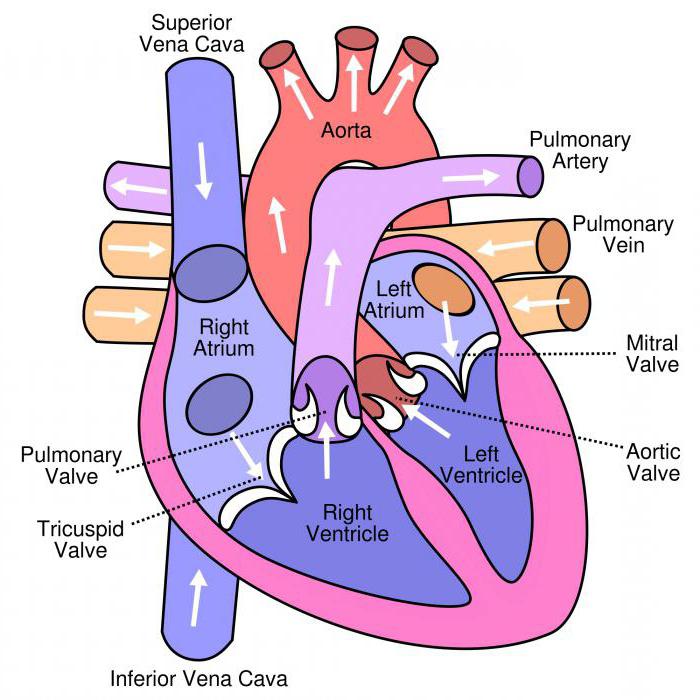
On the left side, this is a bicuspid arterial valve (DAC), and on the right, a tricuspid valve (tricuspid valve). Contraction and relaxation of the heart are called systole and diastole, respectively. Blood is distilled in only one direction: from the upper sections to the ventricles, and from them into the corresponding arteries. So, from its left ventricle, when it is reduced, a portion of blood is ejected with force into the aorta, the blood pressure in it is about 105 mm / RT. Art. A powerful push, in accordance with the laws of physics, quickly spreads in the form of a wave into the system of arteries. We perceive the heart rate as a pulse. It can be well felt on the wrist or carotid arteries.
Liquid tissue in the human body
The functions of the transport of oxygen and nutrients, the removal of toxins and toxins, as well as the production of antibodies are performed by the circulatory system. Blood, the structure of which can be represented as a mixture of cells (leukocytes, erythrocytes and platelets) and plasma (liquid part), provides the fulfillment of the above tasks.
In the human body there are hematopoietic tissues, one of which is myeloid. It is leading in the red bone marrow, located in the diaphysis and contains stem (hematopoietic cells), which are the precursors of red blood cells, white blood cells and platelets.
Features of the structure of blood
The red color of the blood is due to the presence of a hemoglobin pigment. It is he who is responsible for the transport of gases dissolved in the blood - oxygen and carbon monoxide. It can take two forms: oxyhemoglobin and carboxyhemoglobin. Blood plasma is 90% water.
The remaining substances are proteins (albumin, fibrinogen, gamma globulin) and mineral salts, the main of which is sodium chloride. The formed elements of the blood perform such functions:
- red blood cells - carry oxygen;
- white blood cells, or white blood cells (neutrophils, eosinophils, T-lymphocytes, etc.), are involved in the formation of immunity;
- platelets - help stop bleeding in violation of the integrity of the walls of blood vessels (responsible for blood coagulation).
The human circulatory system, thanks to the various functions of the blood, is essential in maintaining homeostasis of the body.
Body vessels: arteries, veins, capillaries
To understand what organs the human circulatory system consists of, you need to imagine it as a network of tubes having different diameters and wall thicknesses. Arteries have a powerful muscle wall, as the blood moves through them with high speed and high pressure. Therefore, arterial bleeding is very dangerous, as a result of which a person loses a large amount of blood in a short time. This can be fatal.
The veins have soft walls, abundantly equipped with lunar valves. They provide the movement of blood in the vessels in only one direction - to the main muscle organ of the circulatory system. Since venous blood is forced to overcome the force of gravity in order to rise to the heart, and the pressure in the veins is low, these valves do not allow blood to move back, that is, from the heart.
A network of capillaries with a microscopic diameter of the walls performs the main function of gas exchange. It is in them that carbon dioxide (carbon dioxide) and toxins from tissue cells enter, and capillary blood, in turn, gives oxygen to the cells necessary for their vital functions. In total, there are more than 150 billion capillaries in the body, the total length of which in an adult is about 100 thousand km.
A special functional adaptation of the human body, which provides a constant supply of organs and tissues with necessary substances, is collateral circulation, which can be observed both in physiologically normal conditions and in complex disorders of the system (for example, a clogged vessel).
Large circle of blood circulation
Let us return to the question of what organs the human circulatory system consists of. Recall that the vicious circle of blood circulation, discovered by Harvey, originates in the left ventricle and ends in the right atrium.
The aorta, as the main artery in the body and the beginning of a large circle of blood circulation, removes oxygen-enriched blood from the left ventricle. Through a system of vessels extending from the aorta and branching throughout the human body, blood enters all parts of the body and organs, saturating them with oxygen, performing the functions of metabolism and transport of nutrients.
From the upper body (head, shoulders, chest, upper limbs), venous blood saturated with carbon dioxide is collected in the superior vena cava, and from the lower half of the body into the inferior vena cava. Both vena cava flow into the right atrium, closing a large circle of blood circulation.
Pulmonary circulation
The circulatory system - the heart, circulatory system - are also included in the so-called small (pulmonary) circle of blood circulation. It was he who discovered Miguel Servet in the middle of the 16th century. This circle starts from the right ventricle and ends in the left atrium.
Venous blood through the right atrioventricular opening from the right atrium enters the right ventricle. From it through the pulmonary trunk, and then through the two pulmonary arteries - left and right - it enters the lungs. And despite the fact that these vessels are called arteries, venous blood flows through them. It enters the right and left lungs, in which there are capillaries surrounding the alveoli (pulmonary vesicles, which make up the lung parenchyma). Between oxygen of the alveoli and connective tissue through the thinnest walls of the capillaries, gas exchange occurs. It is in this part of the body that venous blood turns into arterial blood. Then it enters the post-capillary venules, which are enlarged to 4 pulmonary veins. Through them, arterial blood enters the left atrium, where the pulmonary circulation ends.
Blood circulation in all vessels occurs simultaneously, without stopping or interrupting for even a second.
Coronary circulation
What is an autonomous circulatory system, what organs it consists of, and what are the features of its functioning, have been studied by such scientists as Shumlyansky, Bowman, Gis. They found that coronary or coronary blood circulation, which is carried out by special blood vessels surrounding the heart and extending from the aorta, is of the greatest importance in this system. These are such vessels as the left coronary artery with the main branches, namely: the anterior interventricular, the enveloping branch and the atrial branches. And also it is the right coronary artery with such branches: the right coronary and posterior interventricular.
Blood without oxygen returns back to the muscular organ in three ways: through the coronary sinus, veins entering the atrial cavity, and the smallest vascular branches that flow into the right half of the heart, without even appearing on its epicardium.
Portal vein circle
Since the circulatory system and the organs of the portal vein circle are very important in ensuring the internal constancy of the environment, natural scientists studied in the process of examining a large circle of blood circulation. It was found that from the gastrointestinal tract, spleen and pancreas, blood accumulates in the lower and upper mesenteric veins, which subsequently, when combined, form the portal (portal vein).
The portal vein along with the hepatic artery enters the gate of the liver. Arterial and venous blood in hepatocytes (liver cells) is thoroughly cleaned and then passes through the inferior vena cava into the right atrium. Thus, the purification of blood occurs due to the barrier function of the liver, which provides the circulatory system.
What organs does the auxiliary system consist of?
The composition of the auxiliary organs includes red bone marrow, spleen and the aforementioned liver. Since blood cells do not live for long, approximately 60–90 days, it becomes necessary to dispose of old used blood cells and the synthesis of young ones. It is these processes that provide the auxiliary organs of the circulatory system.
In the red bone marrow containing myeloid tissue, precursors of shaped elements are synthesized.
The spleen, in addition to the function of depositing part of the blood that is not used in the blood circulation, destroys old red blood cells and partially makes up for their loss.
The liver also utilizes dead white blood cells, red blood cells and platelets and stores blood that is not currently involved in the circulatory system.
The circulatory system, which organs it consists of and what functions it performs in the human body, was examined in detail in the article.