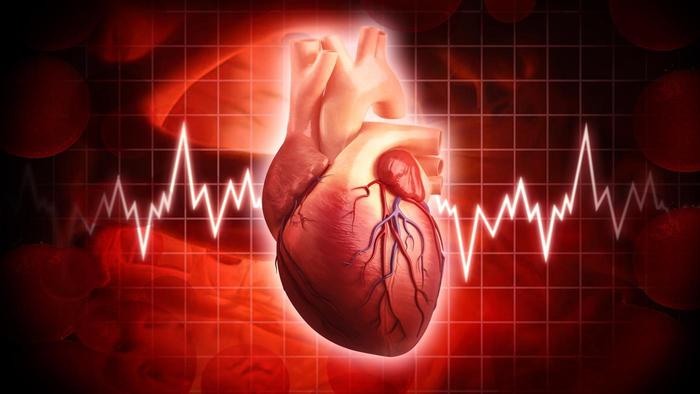
Timely diagnosis of heart disease helps reduce the risk of heart abnormalities. An ECG (electrocardiogram) is the simplest and most informative way to study the functioning of the heart. Data on its abbreviations from the sensors are transmitted to the paper in the form of a graphic image, which consists of lines and teeth of various shapes and heights. The results are used to make a diagnosis. Doing ECG decoding on your own does not make sense. However, in order to have a general idea, it’s good to know in general terms what the cardiogram shows.
How to decrypt a cardiogram?
Decryption is done by a cardiologist. He checks the patency of electrical impulses, the presence of inflammatory processes in the heart muscle, electrolyte metabolic disturbances. Data on heart contractions on an ECG are displayed as:
- Prongs - they can be directed up and are considered positive or down and be negative.
- Segments - the distance between adjacent teeth.
- Intervals - intervals including one tooth and a segment.
To collect information, twelve leads are used: three are called standard and are designated using Roman numerals - I, II, III; three - unipolar reinforced from the lower extremities; six unipolar reinforced thoracic. In case of severe arrhythmia and an abnormally located heart, additional thoracic D, A, I are used.
To decrypt the ECG, you need to know the time period that corresponds to one cell on paper. According to the standard, its value of 1 mm corresponds to 0.04 seconds, at a speed of 25 mm / sec. The intervals from one R wave to the other determine the rhythm of the contraction of the heart. Having information about the speed of data recording and determining the number of cells between the R waves, calculate the heart rate, the norm of which is in the range from 60 to 80 beats per minute. P wave determines excitations in the heart muscle. Sinus rhythm confirms the normal functioning of the heart of a healthy person. One of the essential indicators is the displacement of the electrical axis of the heart. A sharp shift is characteristic of the pathology of the cardiovascular system. Of great importance is the evaluation of segments on the ECG. The largest of them are: P - Q, S - T. The analysis takes into account their length and rise above the horizontal axis. The duration of the site depends on the heart rate and means normal heart function or rhythm disturbance.
ECG reading
When reading an electrocardiogram, the teeth are evaluated by the magnitude of the amplitude of the oscillations, vertically and their width, horizontally. Each tooth is marked with a Latin letter and reflects how an electric current pulse passes through a conditioned area of the heart. It turns out that the tooth:
- P is the reaction of the atria to an electrical impulse. In a healthy person, the tooth is positive, up to 2.5 mm high, the apex is rounded, and the duration is no more than 0.1 s. An abnormality from the ECG norm is the acute configuration of the tooth, resulting from hypertrophy of one of the atria and bifurcation of the apex.
- Q - indicates the passage of the impulse along the septum between the ventricles. During normal operation of the organ, it is negative, the duration is possible up to 0.03 s. In children, it can deeply sink, which is not a pathology.
- R - the main tooth, reflecting the depolarization of the ventricles of the myocardium. By the magnitude of the fluctuations, this is the highest tooth, and the horizontal length does not exceed the value of Q.
- S - the end of excitement in the ventricles of the heart. It is negative, up to 2 mm deep.
- T - shows how the potential of muscle tissue of the heart is restored. Normally, on an ECG in adults, it is above the electrical axis, and its height is one third of the R wave. The shape of the vertex is smoothed, and the duration is from 0.16 to 2.4 s. A large amplitude indicates vegetative pathologies of heart activity. The negative value of the tooth in the form of an isosceles triangle is a sign of myocardial infarction.
- U - on the cardiogram occurs with hypokalemia, hypercalcemia, as well as in athletes after intensive training.
Decoding of the electrocardiogram
Of particular importance in decoding an electrocardiogram is the rhythm. Sinus rhythm is considered the norm on the ECG, pathology - all other types.
Features on the cardiogram of various rhythms:
- Normal. On the printout of the heart in lead II, the P wave is present in front of each QRS complex and is characterized by a positive value. It is noted that all P waves on one lead must be of the same length, shape and width.
- Atrial P waves in the II and III standard leads take negative values and are located in front of each QRS complex.
- Atrioventricular. On a paper carrier of the research results, either the absence of P waves is observed, or they appear after the QRS complex. 40-60 heartbeats occur per minute.
- Ventricular. It is characterized by a significant increase in the width of the QRS complex and the absence of the correct sequence: P wave and QRS complex. The heart contracts less than 40 times per minute.
Detailed decoding of the electrocardiogram
For a strict decoding, the teeth are analyzed, and their area is calculated, and additional leads are used. In everyday practice, the electric axis is often used. Any individual has certain features in the structure of the chest, possibly an unusual arrangement of the heart, different ventricular masses and unequal conductivity in them - all this affects the ECG. As a result, in conclusion, both the vertical and horizontal directions along the electric axis are described. It should be noted that decryption is always carried out in the prescribed manner, this helps to differentiate normal indicators from detected anomalies. The following sequence applies:
- The heart rhythm is analyzed, the heart rate (HR) is calculated. ECG norm options: sinus rhythm, heart rate 60–80 beats per minute.
- The intervals of the ventricular contraction phase are calculated. Normal QT values are less than or equal to 0.4 s. When the interval is lengthened, there is a suspicion of myocarditis, atherosclerosis, rheumatism and coronary heart disease, shortened - it can be caused by hypercalcemia.
- EOS (electrical axis of the heart) - calculated by contour. The height of the R and S teeth is taken as the basis. According to the norm, it is believed that R is always higher than S. Otherwise, there is a high probability of disturbances in the work of the ventricles.
- QRS - explore the width of this complex. Normally, on an ECG in adults, it is 0.3 s. With other values, they speak of ventricular hypertrophy.
- ST - the segment is normally located on the contour. The T wave is positive and asymmetric.
A full decryption is carried out by a cardiologist or ambulance.
Why do ECG readings differ in one patient?
Two ECG results obtained one after another can have different values. The reason for this phenomenon lies in the following factors:
- inaccurate gluing of the cardiogram;
- incorrect reading of Roman numerals;
- loss when cutting data on the first P wave or the last T;
- the influence of nearby electrical appliances on the values of alternating current in the network;
- uncomfortable position of the patient or his excitement before the procedure;
- electrode misalignment or incorrect placement.
It should be borne in mind that the most accurate results are obtained on a stationary electrocardiograph.
Norms of electrocardiograms in adults
The norm of the ECG of the heart is as follows:
- The rhythm of heart contractions is sinus.
- QT - the total duration of ventricular contraction does not exceed 0.4 s.
- The pulse per minute is 60–80 beats. It is determined by the length of the gap between the R teeth and is considered normal when the difference in their length is not more than 10 percent.
- The tooth width P has a reading of 0.1 s.
- The length of the interval PQ the passage of the pulse through the atrioventricular node is 0.1 s.
- QRS - a set of teeth, shows the excitation of the ventricles and is 0.3 s.
- T - the presence of this tooth reflects the relaxation and restoration of the ventricles of the myocardium.
- RR - the average value is 0.6 s.
For any deviations from the norms, the doctor makes a conclusion about violations in the body.
Diseases of the cardiovascular system that can be determined by ECG
It turns out that the onset of some ailments, as well as malignant and benign tumors, congenital heart defects and a defective state of the vessels cannot be determined by electrography.
What diseases does the ECG show? Having decrypted the data from paper media, a cardiologist can identify the following ailments in a patient:
- Arrhythmia is a violation of the formation and promotion of an impulse with a failure of the rhythm. An increase in the gap R - R occurs and fluctuations in the intervals P - Q and Q - T are noted.
- Tachycardia - significantly reduced heart muscle contraction. The intervals between segments are reduced, RS - T is slightly shifted.
- Angina pectoris - with the disease, pain occurs in the region of the heart, and the amplitude of the T wave changes on the ECG and depression of the S-T segment appears.
- Bradycardia - a slowing of the heart occurs. Unlike the norm on the ECG, a decrease in the heart rhythm is visible, the gap between the segments increases, the amplitude of the teeth changes.
- Extrasystole - characterized by rhythm disturbance. There is a deformation of the set of QRS teeth and the absence of P wave, extrasystoles are changed.
- Myocardial infarction - the R wave is absent on electrocardiography, T has a negative value, the S - T segment is located above the isoline.
The norm of indicators of a cardiogram in children
Normal ECG in children are the following values:
- The heart rate of a healthy child is sinus.
- The width of the tooth P is not more than 0.1 s.
- The pulse in children under three years old should not exceed 110 beats per minute, up to five years of age no more than 100, and in adolescents no more than 90.
- PQ - the indicator varies depending on age and matters: under 14 years old - 0.16 s; at the age of 14-17 years - 0.18 s; and older 1.7–0.2 s.
- QRS - in all children, regardless of age category, is 0.06–0.1 s.
- the norm of the QT interval on the ECG should not exceed a value of 0.4 s.
Some abnormalities in children found on the electrocardiogram go away with age. The doctor will explain the situation to the parents or prescribe the appropriate treatment.
Deciphering an electrocardiogram for a heart attack
The most serious diagnosis that can be determined using an ECG is myocardial infarction. It reveals necrotic areas, determines the location, and establishes the difference between acute heart attack from scars and aneurysms that arose with other diseases. On the ECG, there are certain signs by which various stages of the disease are diagnosed:
- Acute - lasts from three hours to three days from the onset of circulatory disorders. In contrast to the ECG norm, the following changes occur: the Q wave may not exist at all; in its presence, the R wave has a small amplitude or is also absent. In this case, a Q - S segment appears, which indicates a transmural infarction. The second sign of an acute heart attack is a rise of at least 4 mm above the isoline of the S - T segment with the formation of one large T wave.
- Acute - lasts from two to three weeks, the Q wave is visible on the electrocardiogram - wide and with large amplitude and the T wave with a negative value.
- Subacute - its duration is up to three months. The ECG differs from the norm by a large negative T wave, in some cases, by an elevation of the S - T segment, which signals a cardiac aneurysm.
- Cicatricial - the final stage, at the site of damage, connective tissue is formed, which is not able to contract. The appearance of a scar on the ECG is marked by a Q wave, which remains on the cardiogram constantly. The T wave remains negative, has a small amplitude.
Electrocardiogram changes after exercise
Electrocardiography has been used in medicine for more than a hundred years. Many heart diseases are diagnosed precisely thanks to the ECG. But there are pathologies that are very difficult to identify, because the patient goes to the doctor when he is in remission. Therefore, in some cases, electrocardiography is performed during and after exercise. To do this, use:
- functional tests;
- bicycle ergometer;
- American method - treadmill test;
- Holter monitoring.
When removing the cardiogram after loading, it is noted that the P wave is located above the horizontal axis and is positive, has a pointed shape. The PQ interval is short, but the load does not affect the QRS complex. S - T segment becomes omitted. It is noted that with increasing load, depression (lowering) of S - T increases. In people with a healthy heart, an ECG after loading, the norm of the S - T segment is the oblique form. The amplitude of the T wave decreases, and it takes a flattened form, which is not considered a sign of coronary insufficiency.
Chronic heart failure (CHF)
CHF occurs with the progression of structural and functional changes in the heart, when it is not able to throw out the necessary amount of oxygen-containing blood for all organs and tissues. It occurs when the myocardium is damaged by infections, injuries, cardiotoxic drugs, or with an increase in vascular resistance due to cardiosclerosis and valve defects. The inability of the heart, like a pump, to pump the necessary amount of blood into the vessels, causes oxygen starvation of organs and the inability to remove all toxic substances from the body. All systems suffer to varying degrees from this. For a long period, the heart copes with chronic failure, increasing the number of contractions and muscle mass of the heart to enhance work.
The individual feels normal at this time. But once the mechanism is exhausted, and complaints about feeling unwell appear. The patient seeks medical help, and the doctor prescribes the study. Will ECG show heart failure? For its diagnosis, of course, ECG indicators are also used, although it does not reveal any specific signs, and at the early stage of the disease in the absence of symptoms, the electrocardiogram remains normal. With the development of the disease, it allows you to use the graph to make an assessment of changes in the size and rhythm of the heart, and can also provide information about previously transferred diseases. In addition, with the help of an ECG, the effectiveness of drug therapy is monitored.
Conclusion
Decoding ECG broken lines and their angles to the horizontal axis is a complex process that requires special medical knowledge and the ability to put them into practice. The readings of the electrocardiogram are affected not only by the general state of health of the individual and the work of the heart muscle, but also by other processes that occur in the body. A cardiologist should know not only the ECG parameters of a healthy person, but also options for deviations that are not always considered anomalies.